V. Rajinikanth
Harmony-Search and Otsu based System for Coronavirus Disease Detection using Lung CT Scan Images
Apr 06, 2020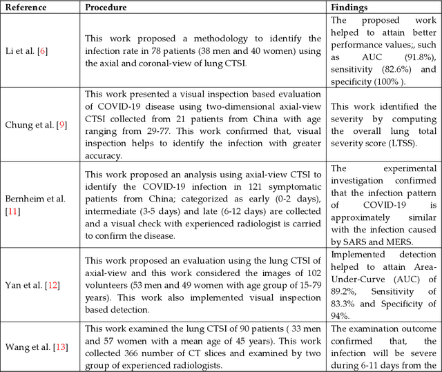
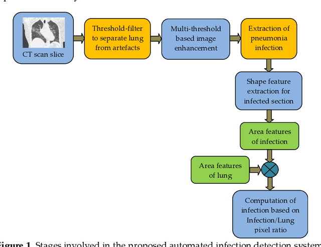
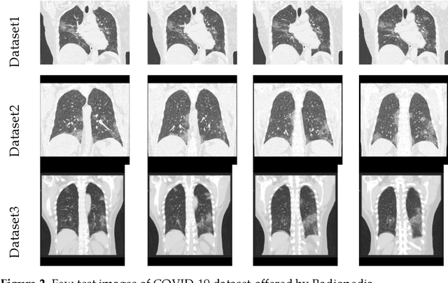
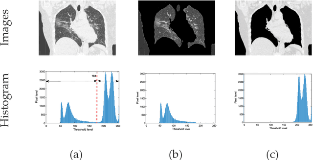
Abstract:Pneumonia is one of the foremost lung diseases and untreated pneumonia will lead to serious threats for all age groups. The proposed work aims to extract and evaluate the Coronavirus disease (COVID-19) caused pneumonia infection in lung using CT scans. We propose an image-assisted system to extract COVID-19 infected sections from lung CT scans (coronal view). It includes following steps: (i) Threshold filter to extract the lung region by eliminating possible artifacts; (ii) Image enhancement using Harmony-Search-Optimization and Otsu thresholding; (iii) Image segmentation to extract infected region(s); and (iv) Region-of-interest (ROI) extraction (features) from binary image to compute level of severity. The features that are extracted from ROI are then employed to identify the pixel ratio between the lung and infection sections to identify infection level of severity. The primary objective of the tool is to assist the pulmonologist not only to detect but also to help plan treatment process. As a consequence, for mass screening processing, it will help prevent diagnostic burden.
Implementation of Deep Neural Networks to Classify EEG Signals using Gramian Angular Summation Field for Epilepsy Diagnosis
Mar 08, 2020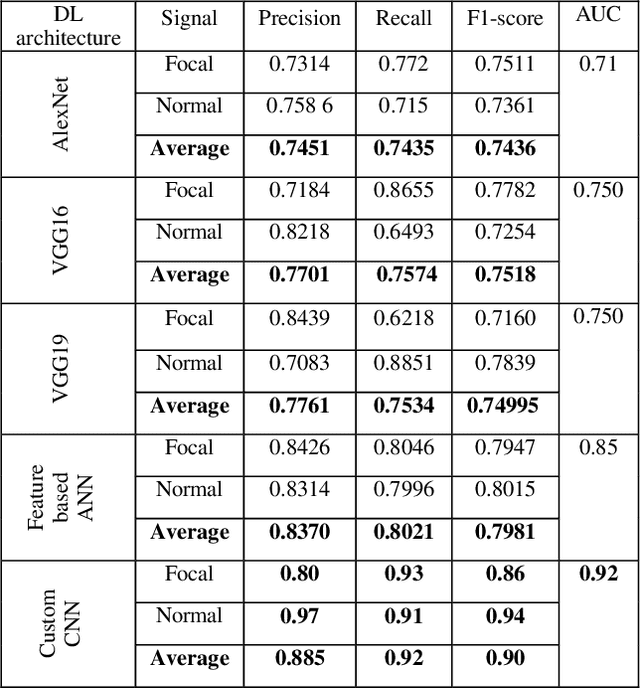
Abstract:This paper evaluates the approach of imaging timeseries data such as EEG in the diagnosis of epilepsy through Deep Neural Network (DNN). EEG signal is transformed into an RGB image using Gramian Angular Summation Field (GASF). Many such EEG epochs are transformed into GASF images for the normal and focal EEG signals. Then, some of the widely used Deep Neural Networks for image classification problems are used here to detect the focal GASF images. Three pre-trained DNN such as the AlexNet, VGG16, and VGG19 are validated for epilepsy detection based on the transfer learning approach. Furthermore, the textural features are extracted from GASF images, and prominent features are selected for a multilayer Artificial Neural Network (ANN) classifier. Lastly, a Custom Convolutional Neural Network (CNN) with three CNN layers, Batch Normalization, Max-pooling layer, and Dense layers, is proposed for epilepsy diagnosis from GASF images. The results of this paper show that the Custom CNN model was able to discriminate against the focal and normal GASF images with an average peak Precision of 0.885, Recall of 0.92, and F1-score of 0.90. Moreover, the Area Under the Curve (AUC) value of the Receiver Operating Characteristic (ROC) curve is 0.92 for the Custom CNN model. This paper suggests that Deep Learning methods widely used in image classification problems can be an alternative approach for epilepsy detection from EEG signals through GASF images.
 Add to Chrome
 Add to Firefox
 Add to Edge