Takayuki Obata
Applied MRI Research, Department of Molecular imaging and Theranostics, National Institute of Radiological Sciences, QST, Department of Molecular imaging and Theranostics, National Institute of Radiological Sciences, QST
Oscillating-gradient spin-echo diffusion-weighted imaging (OGSE-DWI) with a limited number of oscillations: II. Asymptotics
Sep 14, 2023Abstract:Oscillating-gradient spin-echo diffusion-weighted magnetic resonance imaging (OGSE-DWI) has been promoted as a promising technique for studying the microstructure of complex hydrated matter in the frequency domain. The target of the OGSE-DWI technique is the spectral density of molecular diffusion, $u_{2}(\omega)$, which is predicted to obey a set of asymptotic universality relations that are linked to the global organisation of the sample. So, in principle the complex microstructure of a medium can be classified by measuring the spectral density in its low- and high-frequency limits. However, due to practical limitations on the spectral resolution and range of the technique, it is not possible to directly sample the spectral density with OGSE-DWI. Rather, information about the spectral density can be obtained only indirectly through the quantities $U_{kk}$ & $U_{k0}$, which are filtered representations of $u_{2}(\omega)$. The purpose of this study is to investigate how the universal behaviour of $u_{2}(\omega)$ emerges in the asymptotic behaviour of OGSE-DWI signal.
A neural network model that learns differences in diagnosis strategies among radiologists has an improved area under the curve for aneurysm status classification in magnetic resonance angiography image series
Feb 03, 2020


Abstract:Purpose: To construct a neural network model that can learn the different diagnosing strategies of radiologists to better classify aneurysm status in magnetic resonance angiography images. Materials and methods: This retrospective study included 3423 time-of-flight brain magnetic resonance angiography image series (subjects: male 1843 [mean age, 50.2 +/- 11.7 years], female 1580 [50.8 +/- 11.3 years]) recorded from November 2017 through January 2019. The image series were read independently for aneurysm status by one of four board-certified radiologists, who were assisted by an established deep learning-based computer-assisted diagnosis (CAD) system. The constructed neural networks were trained to classify the aneurysm status of zero to five aneurysm-suspicious areas suggested by the CAD system for each image series, and any additional aneurysm areas added by the radiologists, and this classification was compared with the judgment of the annotating radiologist. Image series were randomly allocated to training and testing data in an 8:2 ratio. The accuracy of the classification was compared by receiver operating characteristic analysis between the control model that accepted only image data as input and the proposed model that additionally accepted the information of who the annotating radiologist was. The DeLong test was used to compare areas under the curves (P < 0.05 was considered significant). Results: The area under the curve was larger in the proposed model (0.845) than in the control model (0.793), and the difference was significant (P < 0.0001). Conclusion: The proposed model improved classification accuracy by learning the diagnosis strategies of individual annotating radiologists.
The utility of a convolutional neural network for generating a myelin volume index map from rapid simultaneous relaxometry imaging
Apr 24, 2019


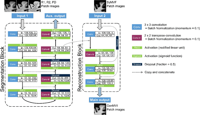
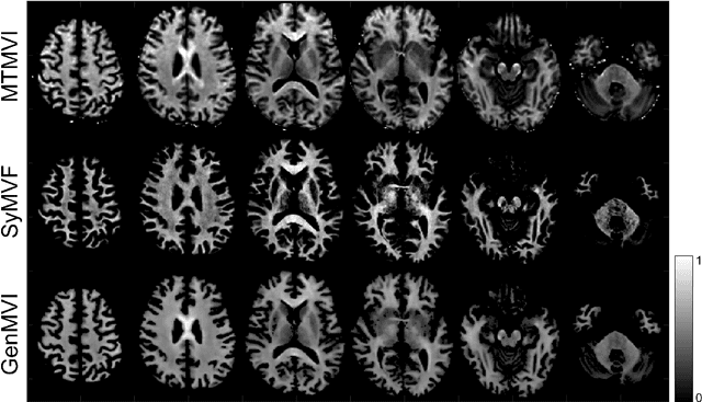
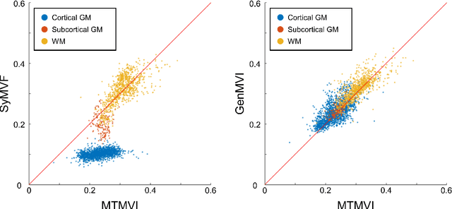
Abstract:Background and Purpose: A current algorithm to obtain a synthetic myelin volume fraction map (SyMVF) from rapid simultaneous relaxometry imaging (RSRI) has a potential problem, that it does not incorporate information from surrounding pixels. The purpose of this study was to develop a method that utilizes a convolutional neural network (CNN) to overcome this problem. Methods: RSRI and magnetization transfer images from 20 healthy volunteers were included. A CNN was trained to reconstruct RSRI-related metric maps into a myelin volume-related index (generated myelin volume index: GenMVI) map using the myelin volume index map calculated from magnetization transfer images (MTMVI) as reference. The SyMVF and GenMVI maps were statistically compared by testing how well they correlated with the MTMVI map. The correlations were evaluated based on: (i) averaged values obtained from 164 atlas-based ROIs, and (ii) pixel-based comparison for ROIs defined in four different tissue types (cortical and subcortical gray matter, white matter, and whole brain). Results: For atlas-based ROIs, the overall correlation with the MTMVI map was higher for the GenMVI map than for the SyMVF map. In the pixel-based comparison, correlation with the MTMVI map was stronger for the GenMVI map than for the SyMVF map, and the difference in the distribution for the volunteers was significant (Wilcoxon sign-rank test, P<.001) in all tissue types. Conclusion: The proposed method is useful, as it can incorporate more specific information about local tissue properties than the existing method.
 Add to Chrome
 Add to Firefox
 Add to Edge