Sung-Liang Chen
De-Noising of Photoacoustic Microscopy Images by Deep Learning
Jan 12, 2022


Abstract:As a hybrid imaging technology, photoacoustic microscopy (PAM) imaging suffers from noise due to the maximum permissible exposure of laser intensity, attenuation of ultrasound in the tissue, and the inherent noise of the transducer. De-noising is a post-processing method to reduce noise, and PAM image quality can be recovered. However, previous de-noising techniques usually heavily rely on mathematical priors as well as manually selected parameters, resulting in unsatisfactory and slow de-noising performance for different noisy images, which greatly hinders practical and clinical applications. In this work, we propose a deep learning-based method to remove complex noise from PAM images without mathematical priors and manual selection of settings for different input images. An attention enhanced generative adversarial network is used to extract image features and remove various noises. The proposed method is demonstrated on both synthetic and real datasets, including phantom (leaf veins) and in vivo (mouse ear blood vessels and zebrafish pigment) experiments. The results show that compared with previous PAM de-noising methods, our method exhibits good performance in recovering images qualitatively and quantitatively. In addition, the de-noising speed of 0.016 s is achieved for an image with $256\times256$ pixels. Our approach is effective and practical for the de-noising of PAM images.
Image enhancement in acoustic-resolution photoacoustic microscopy enabled by a novel directional algorithm
Nov 19, 2021


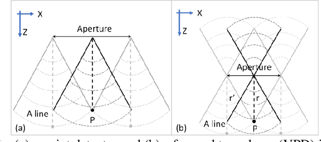
Abstract:Acoustic-resolution photoacoustic microscopy (AR-PAM) is a promising tool for microvascular imaging. In the focal region, resolution of AR-PAM is determined by the ultrasound transducer and ultimately limited by acoustic diffraction. In the out-of-focus region, resolution deteriorates with increasing distance from the focal plane, which restricts depth of focus (DOF). Besides, a trade-off exists between resolution and DOF. Previously, synthetic aperture focusing technique (SAFT) and/or deconvolution methods have been demonstrated to enhance AR-PAM images. However, they suffer from issues in low resolution, low signal-to-noise ratio (SNR), and/or poor image fidelity. Here, we propose a novel algorithm for AR-PAM to enhance image resolution, SNR, and fidelity. The algorithm consists of a Fourier accumulation SAFT (FA-SAFT) and a directional model-based (D-MB) deconvolution method. Inspired from Fourier denoising technique and directional SAFT, FA-SAFT mainly compensates for the defocusing effect. Besides, D-MB deconvolution enhances the resolution as well as preserves the image fidelity, especially for the objects with line patterns such as microvasculature. Full width at half maximum of 26-31 um over DOF of 1.8 mm and minimum resolvable distance of 46-49 um are experimentally achieved by imaging tungsten wire phantom. Moreover, imaging of leaf skeleton phantom and in vivo imaging of mouse blood vessels also prove that our algorithm is capable of providing high-resolution, high-SNR, and good-fidelity results for complex structures and for in vivo applications.
Adaptive Weighting Depth-variant Deconvolution of Fluorescence Microscopy Images with Convolutional Neural Network
Jul 07, 2019


Abstract:Fluorescence microscopy plays an important role in biomedical research. The depth-variant point spread function (PSF) of a fluorescence microscope produces low-quality images especially in the out-of-focus regions of thick specimens. Traditional deconvolution to restore the out-of-focus images is usually insufficient since a depth-invariant PSF is assumed. This article aims at handling fluorescence microscopy images by learning-based depth-variant PSF and reducing artifacts. We propose adaptive weighting depth-variant deconvolution (AWDVD) with defocus level prediction convolutional neural network (DelpNet) to restore the out-of-focus images. Depth-variant PSFs of image patches can be obtained by DelpNet and applied in the afterward deconvolution. AWDVD is adopted for a whole image which is patch-wise deconvolved and appropriately cropped before deconvolution. DelpNet achieves the accuracy of 98.2%, which outperforms the best-ever one using the same microscopy dataset. Image patches of 11 defocus levels after deconvolution are validated with maximum improvement in the peak signal-to-noise ratio and structural similarity index of 6.6 dB and 11%, respectively. The adaptive weighting of the patch-wise deconvolved image can eliminate patch boundary artifacts and improve deconvolved image quality. The proposed method can accurately estimate depth-variant PSF and effectively recover out-of-focus microscopy images. To our acknowledge, this is the first study of handling out-of-focus microscopy images using learning-based depth-variant PSF. Facing one of the most common blurs in fluorescence microscopy, the novel method provides a practical technology to improve the image quality.
 Add to Chrome
 Add to Firefox
 Add to Edge