Stephanie Thorn
Transformer-based Dual-domain Network for Few-view Dedicated Cardiac SPECT Image Reconstructions
Jul 23, 2023


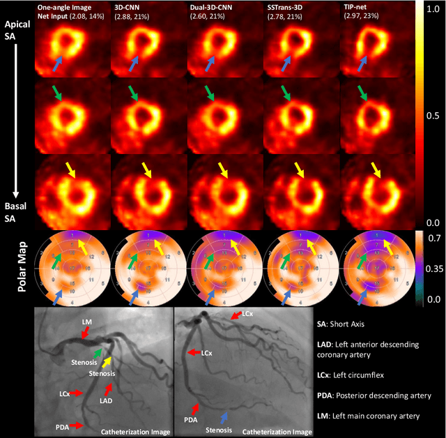
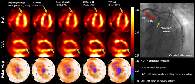
Abstract:Cardiovascular disease (CVD) is the leading cause of death worldwide, and myocardial perfusion imaging using SPECT has been widely used in the diagnosis of CVDs. The GE 530/570c dedicated cardiac SPECT scanners adopt a stationary geometry to simultaneously acquire 19 projections to increase sensitivity and achieve dynamic imaging. However, the limited amount of angular sampling negatively affects image quality. Deep learning methods can be implemented to produce higher-quality images from stationary data. This is essentially a few-view imaging problem. In this work, we propose a novel 3D transformer-based dual-domain network, called TIP-Net, for high-quality 3D cardiac SPECT image reconstructions. Our method aims to first reconstruct 3D cardiac SPECT images directly from projection data without the iterative reconstruction process by proposing a customized projection-to-image domain transformer. Then, given its reconstruction output and the original few-view reconstruction, we further refine the reconstruction using an image-domain reconstruction network. Validated by cardiac catheterization images, diagnostic interpretations from nuclear cardiologists, and defect size quantified by an FDA 510(k)-cleared clinical software, our method produced images with higher cardiac defect contrast on human studies compared with previous baseline methods, potentially enabling high-quality defect visualization using stationary few-view dedicated cardiac SPECT scanners.
 Add to Chrome
 Add to Firefox
 Add to Edge