Stephan W. Anderson
Wireless, Customizable Coaxially-shielded Coils for Magnetic Resonance Imaging
Dec 19, 2023Abstract:Anatomy-specific RF receive coil arrays routinely adopted in magnetic resonance imaging (MRI) for signal acquisition, are commonly burdened by their bulky, fixed, and rigid configurations, which may impose patient discomfort, bothersome positioning, and suboptimal sensitivity in certain situations. Herein, leveraging coaxial cables' inherent flexibility and electric field confining property, for the first time, we present wireless, ultra-lightweight, coaxially-shielded MRI coils achieving a signal-to-noise ratio (SNR) comparable to or surpassing that of commercially available cutting-edge receive coil arrays with the potential for improved patient comfort, ease of implementation, and significantly reduced costs. The proposed coils demonstrate versatility by functioning both independently in form-fitting configurations, closely adapting to relatively small anatomical sites, and collectively by inductively coupling together as metamaterials, allowing for extension of the field-of-view of their coverage to encompass larger anatomical regions without compromising coil sensitivity. The wireless, coaxially-shielded MRI coils reported herein pave the way toward next generation MRI coils.
Wearable Coaxially-shielded Metamaterial for Magnetic Resonance Imaging
Dec 15, 2023Abstract:Recent advancements in metamaterials have yielded the possibility of a wireless solution to improve signal-to-noise ratio (SNR) in magnetic resonance imaging (MRI). Unlike traditional closely packed local coil arrays with rigid designs and numerous components, these lightweight, cost-effective metamaterials eliminate the need for radio frequency (RF) cabling, baluns, adapters, and interfaces. However, their clinical adoption has been limited by their low sensitivity, bulky physical footprint, and limited, specific use cases. Herein, we introduce a wearable metamaterial developed using commercially available coaxial cable, designed for a 3.0 T MRI system. This metamaterial inherits the coaxially-shielded structure of its constituent coaxial cable, effectively containing the electric field within the cable, thereby mitigating the electric coupling to its loading while ensuring safer clinical adoption, lower signal loss, and resistance to frequency shifts. Weighing only 50g, the metamaterial maximizes its sensitivity by conforming to the anatomical region of interest. MRI images acquired using this metamaterial with various pulse sequences demonstrate an up to 2-fold SNR enhancement when compared to a state-of-the-art 16-channel knee coil. This work introduces a novel paradigm for constructing metamaterials in the MRI environment, paving the way for the development of next-generation wireless MRI technology.
Conformal Metamaterials with Active Tunability and Self-adaptivity for Magnetic Resonance Imaging
Sep 29, 2023


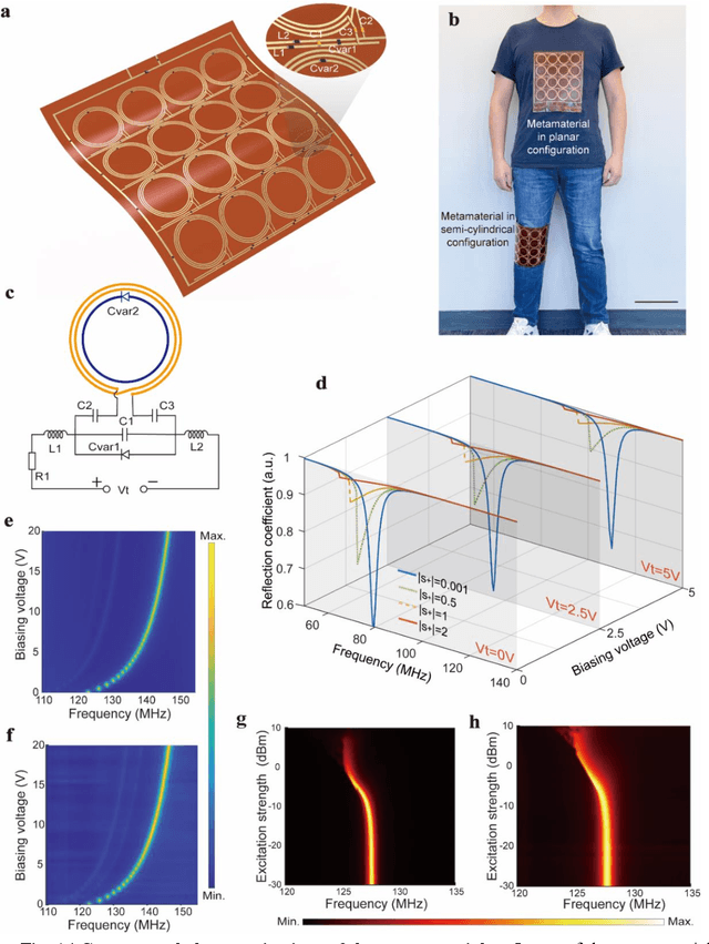
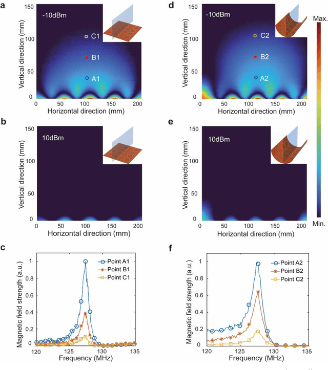
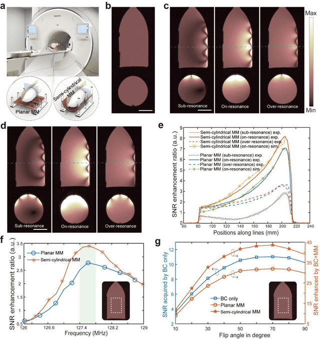
Abstract:Ongoing effort has been devoted to applying metamaterials to boost the imaging performance of magnetic resonance imaging owing to their unique capacity for electromagnetic field confinement and enhancement. However, there are still major obstacles to widespread clinical adoption of conventional metamaterials due to several notable restrictions, namely: their typically bulky and rigid structures, deviations in their optimal resonance frequency, and their inevitable interference with the transmission RF field in MRI. Herein, we address these restrictions and report a conformal, smart metamaterial, which may not only be readily tuned to achieve the desired, precise frequency match with MRI by a controlling circuit, but is also capable of selectively amplifying the magnetic field during the RF reception phase by sensing the excitation signal strength passively, thereby remaining off during the RF transmission phase and thereby ensuring its optimal performance when applied to MRI as an additive technology. By addressing a host of current technological challenges, the metamaterial presented herein paves the way toward the wide-ranging utilization of metamaterials in clinical MRI, thereby translating this promising technology to the MRI bedside.
Attention Hybrid Variational Net for Accelerated MRI Reconstruction
Jun 21, 2023


Abstract:The application of compressed sensing (CS)-enabled data reconstruction for accelerating magnetic resonance imaging (MRI) remains a challenging problem. This is due to the fact that the information lost in k-space from the acceleration mask makes it difficult to reconstruct an image similar to the quality of a fully sampled image. Multiple deep learning-based structures have been proposed for MRI reconstruction using CS, both in the k-space and image domains as well as using unrolled optimization methods. However, the drawback of these structures is that they are not fully utilizing the information from both domains (k-space and image). Herein, we propose a deep learning-based attention hybrid variational network that performs learning in both the k-space and image domain. We evaluate our method on a well-known open-source MRI dataset and a clinical MRI dataset of patients diagnosed with strokes from our institution to demonstrate the performance of our network. In addition to quantitative evaluation, we undertook a blinded comparison of image quality across networks performed by a subspecialty trained radiologist. Overall, we demonstrate that our network achieves a superior performance among others under multiple reconstruction tasks.
 Add to Chrome
 Add to Firefox
 Add to Edge