Soham Mandal
Application of S-Transform on Hyper kurtosis based Modified Duo Histogram Equalized DIC images for Pre-cancer Detection
Apr 30, 2015Abstract:Our proposed hyper kurtosis based histogram equalized DIC images enhances the contrast by preserving the brightness. The evolution and development of precancerous activity among tissues are studied through S-transform (ST). The significant variations of amplitude spectra can be observed due to increased medium roughness from normal tissue were observed in time-frequency domain. The randomness and inhomogeneity of the tissue structures among human normal and different grades of DIC tissues is recognized by ST based timefrequency analysis. This study offers a simpler and better way to recognize the substantial changes among different stages of DIC tissues, which are reflected by spatial information containing within the inhomogeneity structures of different types of tissue.
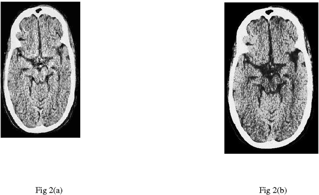
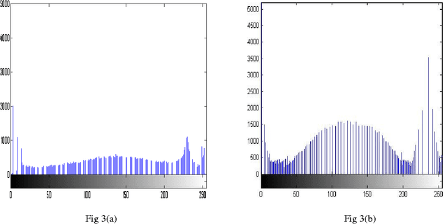
A comparative study between proposed Hyper Kurtosis based Modified Duo-Histogram Equalization (HKMDHE) and Contrast Limited Adaptive Histogram Equalization (CLAHE) for Contrast Enhancement Purpose of Low Contrast Human Brain CT scan images
Apr 07, 2015


Abstract:In this paper, a comparative study between proposed hyper kurtosis based modified duo-histogram equalization (HKMDHE) algorithm and contrast limited adaptive histogram enhancement (CLAHE) has been presented for the implementation of contrast enhancement and brightness preservation of low contrast human brain CT scan images. In HKMDHE algorithm, contrast enhancement is done on the hyper-kurtosis based application. The results are very promising of proposed HKMDHE technique with improved PSNR values and lesser AMMBE values than CLAHE technique.
Wavelet based approach for tissue fractal parameter measurement: Pre cancer detection
Mar 21, 2015

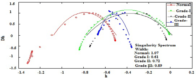
Abstract:In this paper, we have carried out the detail studies of pre-cancer by wavelet coherency and multifractal based detrended fluctuation analysis (MFDFA) on differential interference contrast (DIC) images of stromal region among different grades of pre-cancer tissues. Discrete wavelet transform (DWT) through Daubechies basis has been performed for identifying fluctuations over polynomial trends for clear characterization and differentiation of tissues. Wavelet coherence plots are performed for identifying the level of correlation in time scale plane between normal and various grades of DIC samples. Applying MFDFA on refractive index variations of cervical tissues, we have observed that the values of Hurst exponent (correlation) decreases from healthy (normal) to pre-cancer tissues. The width of singularity spectrum has a sudden degradation at grade-I in comparison of healthy (normal) tissue but later on it increases as cancer progresses from grade-II to grade-III.
Diagnosing Heterogeneous Dynamics for CT Scan Images of Human Brain in Wavelet and MFDFA domain
Mar 12, 2015Abstract:CT scan images of human brain of a particular patient in different cross sections are taken, on which wavelet transform and multi-fractal analysis are applied. The vertical and horizontal unfolding of images are done before analyzing these images. A systematic investigation of de-noised CT scan images of human brain in different cross-sections are carried out through wavelet normalized energy and wavelet semi-log plots, which clearly points out the mismatch between results of vertical and horizontal unfolding. The mismatch of results confirms the heterogeneity in spatial domain. Using the multi-fractal de-trended fluctuation analysis (MFDFA), the mismatch between the values of Hurst exponent and width of singularity spectrum by vertical and horizontal unfolding confirms the same.
 Add to Chrome
 Add to Firefox
 Add to Edge