Shaofeng Yuan
Segmentation of Aortic Vessel Tree in CT Scans with Deep Fully Convolutional Networks
May 16, 2023Abstract:Automatic and accurate segmentation of aortic vessel tree (AVT) in computed tomography (CT) scans is crucial for early detection, diagnosis and prognosis of aortic diseases, such as aneurysms, dissections and stenosis. However, this task remains challenges, due to the complexity of aortic vessel tree and amount of CT angiography data. In this technical report, we use two-stage fully convolutional networks (FCNs) to automatically segment AVT in CTA scans from multiple centers. Specifically, we firstly adopt a 3D FCN with U-shape network architecture to segment AVT in order to produce topology attention and accelerate medical image analysis pipeline. And then another one 3D FCN is trained to segment branches of AVT along the pseudo-centerline of AVT. In the 2023 MICCAI Segmentation of the Aorta (SEG.A.) Challenge , the reported method was evaluated on the public dataset of 56 cases. The resulting Dice Similarity Coefficient (DSC) is 0.920, Jaccard Similarity Coefficient (JSC) is 0.861, Recall is 0.922, and Precision is 0.926 on a 5-fold random split of training and validation set.
CSDN: Combing Shallow and Deep Networks for Accurate Real-time Segmentation of High-definition Intravascular Ultrasound Images
Jan 30, 2023Abstract:Intravascular ultrasound (IVUS) is the preferred modality for capturing real-time and high resolution cross-sectional images of the coronary arteries, and evaluating the stenosis. Accurate and real-time segmentation of IVUS images involves the delineation of lumen and external elastic membrane borders. In this paper, we propose a two-stream framework for efficient segmentation of 60 MHz high resolution IVUS images. It combines shallow and deep networks, namely, CSDN. The shallow network with thick channels focuses to extract low-level details. The deep network with thin channels takes charge of learning high-level semantics. Treating the above information separately enables learning a model to achieve high accuracy and high efficiency for accurate real-time segmentation. To further improve the segmentation performance, mutual guided fusion module is used to enhance and fuse both different types of feature representation. The experimental results show that our CSDN accomplishes a good trade-off between analysis speed and segmentation accuracy.
Extraction of Pulmonary Airway in CT Scans Using Deep Fully Convolutional Networks
Aug 12, 2022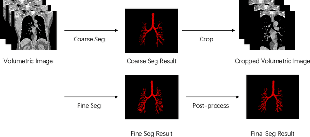


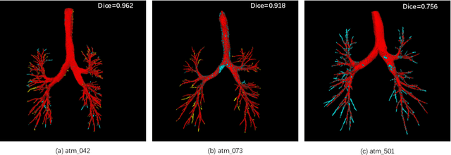
Abstract:Accurate, automatic and complete extraction of pulmonary airway in medical images plays an important role in analyzing thoracic CT volumes such as lung cancer detection, chronic obstructive pulmonary disease (COPD), and bronchoscopic-assisted surgery navigation. However, this task remains challenges, due to the complex tree-like structure of the airways. In this technical report, we use two-stage fully convolutional networks (FCNs) to automatically segment pulmonary airway in thoracic CT scans from multi-sites. Specifically, we firstly adopt a 3D FCN with U-shape network architecture to segment pulmonary airway in a coarse resolution in order to accelerate medical image analysis pipeline. And then another one 3D FCN is trained to segment pulmonary airway in a fine resolution. In the 2022 MICCAI Multi-site Multi-domain Airway Tree Modeling (ATM) Challenge, the reported method was evaluated on the public training set of 300 cases and independent private validation set of 50 cases. The resulting Dice Similarity Coefficient (DSC) is 0.914 $\pm$ 0.040, False Negative Error (FNE) is 0.079 $\pm$ 0.042, and False Positive Error (FPE) is 0.090 $\pm$ 0.066 on independent private validation set.
A Comparison of 1-D and 2-D Deep Convolutional Neural Networks in ECG Classification
Oct 16, 2018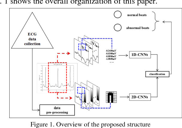


Abstract:Effective detection of arrhythmia is an important task in the remote monitoring of electrocardiogram (ECG). The traditional ECG recognition depends on the judgment of the clinicians' experience, but the results suffer from the probability of human error due to the fatigue. To solve this problem, an ECG signal classification method based on the images is presented to classify ECG signals into normal and abnormal beats by using two-dimensional convolutional neural networks (2D-CNNs). First, we compare the accuracy and robustness between one-dimensional ECG signal input method and two-dimensional image input method in AlexNet network. Then, in order to alleviate the overfitting problem in two-dimensional network, we initialize AlexNet-like network with weights trained on ImageNet, to fit the training ECG images and fine-tune the model, and to further improve the accuracy and robustness of ECG classification. The performance evaluated on the MIT-BIH arrhythmia database demonstrates that the proposed method can achieve the accuracy of 98% and maintain high accuracy within SNR range from 20 dB to 35 dB. The experiment shows that the 2D-CNNs initialized with AlexNet weights performs better than one-dimensional signal method without a large-scale dataset.
A Multi-stage Framework with Context Information Fusion Structure for Skin Lesion Segmentation
Oct 16, 2018
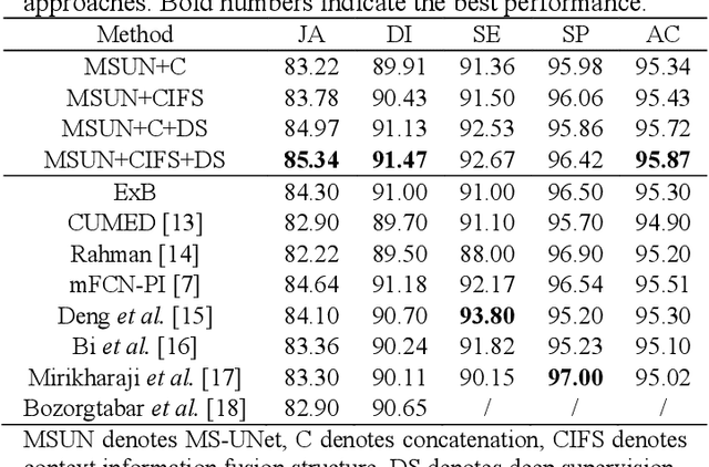

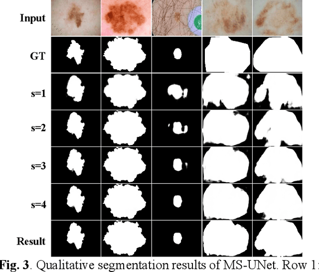
Abstract:The computer-aided diagnosis (CAD) systems can highly improve the reliability and efficiency of melanoma recognition. As a crucial step of CAD, skin lesion segmentation has the unsatisfactory accuracy in existing methods due to large variability in lesion appearance and artifacts. In this work, we propose a framework employing multi-stage UNets (MS-UNet) in the auto-context scheme to segment skin lesion accurately end-to-end. We apply two approaches to boost the performance of MS-UNet. First, UNet is coupled with a context information fusion structure (CIFS) to integrate the low-level and context information in the multi-scale feature space. Second, to alleviate the gradient vanishing problem, we use deep supervision mechanism through supervising MS-UNet by minimizing a weighted Jaccard distance loss function. Four out of five commonly used performance metrics, including Jaccard index and Dice coefficient, show that our approach outperforms the state-ofthe-art deep learning based methods on the ISBI 2016 Skin Lesion Challenge dataset.
 Add to Chrome
 Add to Firefox
 Add to Edge