Reza A. Zoroofi
Analysis of Macula on Color Fundus Images Using Heightmap Reconstruction Through Deep Learning
Dec 28, 2020


Abstract:For medical diagnosis based on retinal images, a clear understanding of 3D structure is often required but due to the 2D nature of images captured, we cannot infer that information. However, by utilizing 3D reconstruction methods, we can recover the height information of the macula area on a fundus image which can be helpful for diagnosis and screening of macular disorders. Recent approaches have used shading information for heightmap prediction but their output was not accurate since they ignored the dependency between nearby pixels and only utilized shading information. Additionally, other methods were dependent on the availability of more than one image of the retina which is not available in practice. In this paper, motivated by the success of Conditional Generative Adversarial Networks(cGANs) and deeply supervised networks, we propose a novel architecture for the generator which enhances the details and the quality of output by progressive refinement and the use of deep supervision to reconstruct the height information of macula on a color fundus image. Comparisons on our own dataset illustrate that the proposed method outperforms all of the state-of-the-art methods in image translation and medical image translation on this particular task. Additionally, perceptual studies also indicate that the proposed method can provide additional information for ophthalmologists for diagnosis.
Heightmap Reconstruction of Macula on Color Fundus Images Using Conditional Generative Adversarial Networks
Sep 05, 2020


Abstract:For medical diagnosis based on retinal images, a clear understanding of 3D structure is often required but due to the 2D nature of images captured, we cannot infer that information. However, by utilizing 3D reconstruction methods, we can construct the 3D structure of the macula area on fundus images which can be helpful for diagnosis and screening of macular disorders. Recent approaches have used shading information for 3D reconstruction or heightmap prediction but their output was not accurate since they ignored the dependency between nearby pixels. Additionally, other methods were dependent on the availability of more than one image of the eye which is not available in practice. In this paper, we use conditional generative adversarial networks (cGANs) to generate images that contain height information of the macula area on a fundus image. Results using our dataset show a 0.6077 improvement in Structural Similarity Index (SSIM) and 0.071 improvements in Mean Squared Error (MSE) metric over Shape from Shading (SFS) method. Additionally, Qualitative studies also indicate that our method outperforms recent approaches.
Region-based Convolution Neural Network Approach for Accurate Segmentation of Pelvic Radiograph
Oct 29, 2019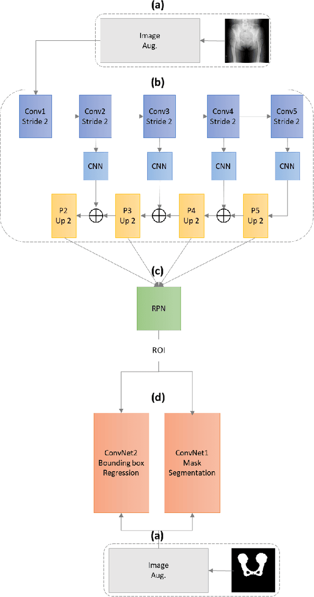
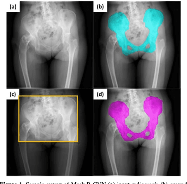
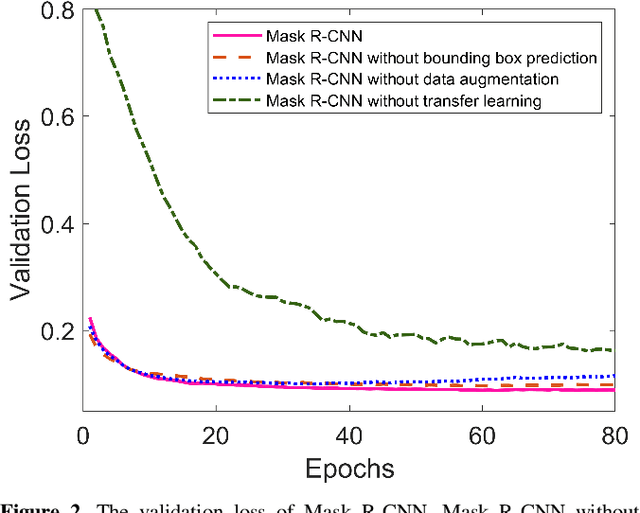
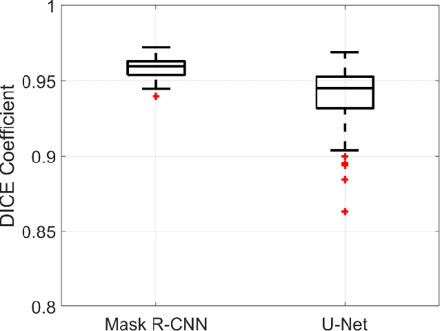
Abstract:With the increasing usage of radiograph images as a most common medical imaging system for diagnosis, treatment planning, and clinical studies, it is increasingly becoming a vital factor to use machine learning-based systems to provide reliable information for surgical pre-planning. Segmentation of pelvic bone in radiograph images is a critical preprocessing step for some applications such as automatic pose estimation and disease detection. However, the encoder-decoder style network known as U-Net has demonstrated limited results due to the challenging complexity of the pelvic shapes, especially in severe patients. In this paper, we propose a novel multi-task segmentation method based on Mask R-CNN architecture. For training, the network weights were initialized by large non-medical dataset and fine-tuned with radiograph images. Furthermore, in the training process, augmented data was generated to improve network performance. Our experiments show that Mask R-CNN utilizing multi-task learning, transfer learning, and data augmentation techniques achieve 0.96 DICE coefficient, which significantly outperforms the U-Net. Notably, for a fair comparison, the same transfer learning and data augmentation techniques have been used for U-net training.
Estimation of Pelvic Sagittal Inclination from Anteroposterior Radiograph Using Convolutional Neural Networks: Proof-of-Concept Study
Oct 26, 2019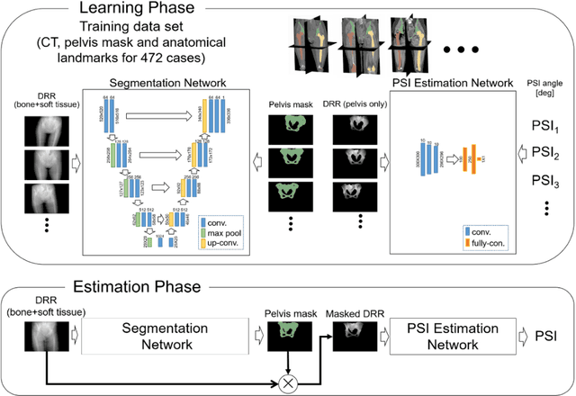
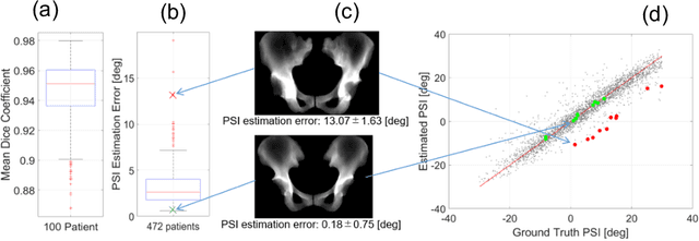
Abstract:Alignment of the bones in standing position provides useful information in surgical planning. In total hip arthroplasty (THA), pelvic sagittal inclination (PSI) angle in the standing position is an important factor in planning of cup alignment and has been estimated mainly from radiographs. Previous methods for PSI estimation used a patient-specific CT to create digitally reconstructed radiographs (DRRs) and compare them with the radiograph to estimate relative position between the pelvis and the x-ray detector. In this study, we developed a method that estimates PSI angle from a single anteroposterior radiograph using two convolutional neural networks (CNNs) without requiring the patient-specific CT, which reduces radiation exposure of the patient and opens up the possibility of application in a larger number of hospitals where CT is not acquired in a routine protocol.
 Add to Chrome
 Add to Firefox
 Add to Edge