Reda Alhajj
AutoCOR: Autonomous Condylar Offset Ratio Calculator on TKA-Postoperative Lateral Knee X-ray
Apr 06, 2022Abstract:The postoperative range of motion is one of the crucial factors indicating the outcome of Total Knee Arthroplasty (TKA). Although the correlation between range of knee flexion and posterior condylar offset (PCO) is controversial in the literature, PCO maintains its importance on evaluation of TKA. Due to limitations on PCO measurement, two novel parameters, posterior condylar offset ratio (PCOR) and anterior condylar offset ratio (ACOR), were introduced. Nowadays, the calculation of PCOR and ACOR on plain lateral radiographs is done manually by orthopedic surgeons. In this regard, we developed a software, AutoCOR, to calculate PCOR and ACOR autonomously, utilizing unsupervised machine learning algorithm (k-means clustering) and digital image processing techniques. The software AutoCOR is capable of detecting the anterior/posterior edge points and anterior/posterior cortex of the femoral shaft on true postoperative lateral conventional radiographs. To test the algorithm, 50 postoperative true lateral radiographs from Istanbul Kosuyolu Medipol Hospital Database were used (32 patients). The mean PCOR was 0.984 (SD 0.235) in software results and 0.972 (SD 0.164) in ground truth values. It shows strong and significant correlation between software and ground truth values (Pearson r=0.845 p<0.0001). The mean ACOR was 0.107 (SD 0.092) in software results and 0.107 (SD 0.070) in ground truth values. It shows moderate and significant correlation between software and ground truth values (Spearman's rs=0.519 p=0.0001412). We suggest that AutoCOR is a useful tool that can be used in clinical practice.
Transfer Learning Through Weighted Loss Function and Group Normalization for Vessel Segmentation from Retinal Images
Dec 16, 2020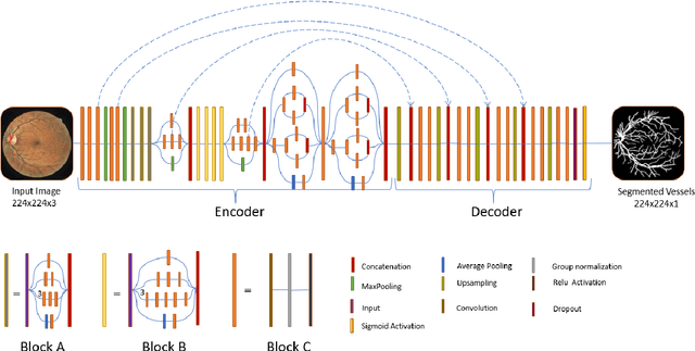
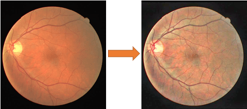
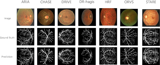

Abstract:The vascular structure of blood vessels is important in diagnosing retinal conditions such as glaucoma and diabetic retinopathy. Accurate segmentation of these vessels can help in detecting retinal objects such as the optic disc and optic cup and hence determine if there are damages to these areas. Moreover, the structure of the vessels can help in diagnosing glaucoma. The rapid development of digital imaging and computer-vision techniques has increased the potential for developing approaches for segmenting retinal vessels. In this paper, we propose an approach for segmenting retinal vessels that uses deep learning along with transfer learning. We adapted the U-Net structure to use a customized InceptionV3 as the encoder and used multiple skip connections to form the decoder. Moreover, we used a weighted loss function to handle the issue of class imbalance in retinal images. Furthermore, we contributed a new dataset to this field. We tested our approach on six publicly available datasets and a newly created dataset. We achieved an average accuracy of 95.60% and a Dice coefficient of 80.98%. The results obtained from comprehensive experiments demonstrate the robustness of our approach to the segmentation of blood vessels in retinal images obtained from different sources. Our approach results in greater segmentation accuracy than other approaches.
Utilizing Transfer Learning and a Customized Loss Function for Optic Disc Segmentation from Retinal Images
Oct 01, 2020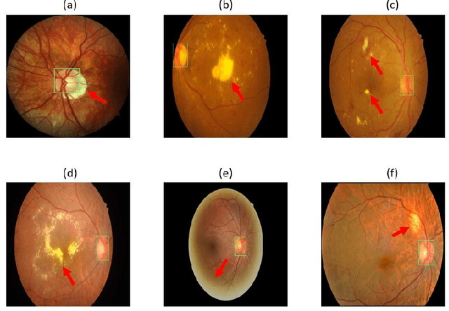
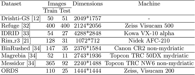


Abstract:Accurate segmentation of the optic disc from a retinal image is vital to extracting retinal features that may be highly correlated with retinal conditions such as glaucoma. In this paper, we propose a deep-learning based approach capable of segmenting the optic disc given a high-precision retinal fundus image. Our approach utilizes a UNET-based model with a VGG16 encoder trained on the ImageNet dataset. This study can be distinguished from other studies in the customization made for the VGG16 model, the diversity of the datasets adopted, the duration of disc segmentation, the loss function utilized, and the number of parameters required to train our model. Our approach was tested on seven publicly available datasets augmented by a dataset from a private clinic that was annotated by two Doctors of Optometry through a web portal built for this purpose. We achieved an accuracy of 99.78\% and a Dice coefficient of 94.73\% for a disc segmentation from a retinal image in 0.03 seconds. The results obtained from comprehensive experiments demonstrate the robustness of our approach to disc segmentation of retinal images obtained from different sources.
A Review on Deep Learning Techniques for the Diagnosis of Novel Coronavirus
Aug 09, 2020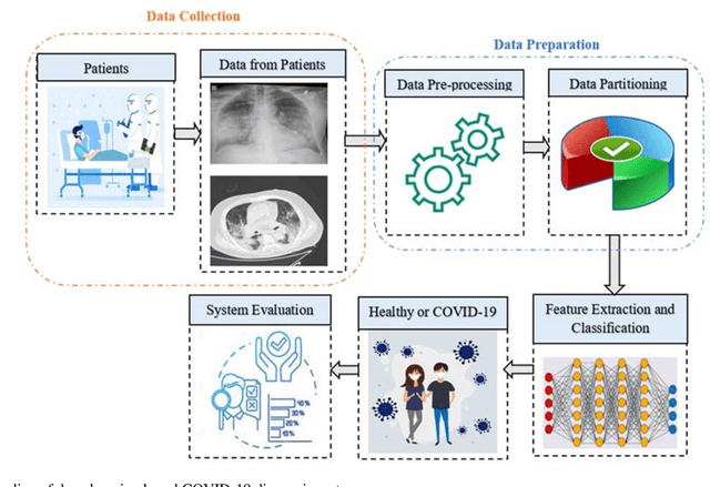
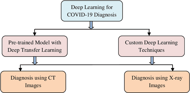
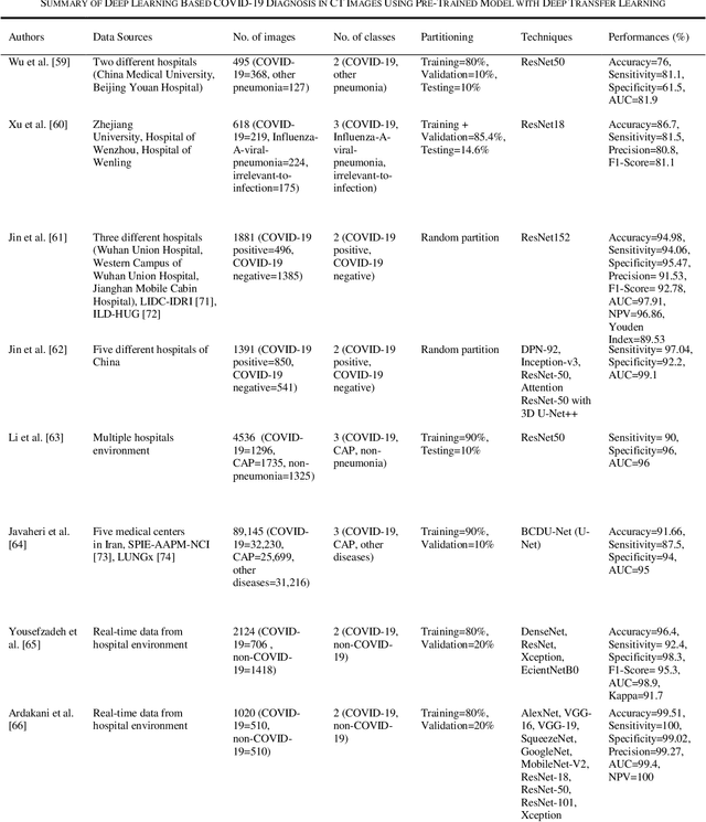
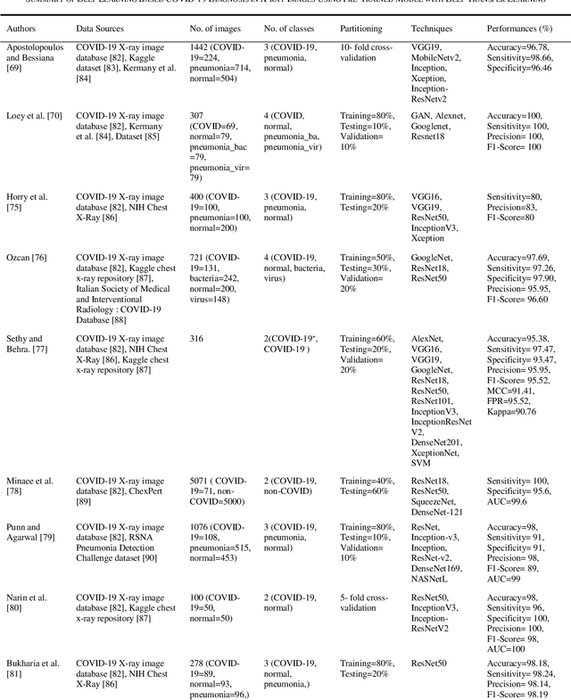
Abstract:Novel coronavirus (COVID-19) outbreak, has raised a calamitous situation all over the world and has become one of the most acute and severe ailments in the past hundred years. The prevalence rate of COVID-19 is rapidly rising every day throughout the globe. Although no vaccines for this pandemic have been discovered yet, deep learning techniques proved themselves to be a powerful tool in the arsenal used by clinicians for the automatic diagnosis of COVID-19. This paper aims to overview the recently developed systems based on deep learning techniques using different medical imaging modalities like Computer Tomography (CT) and X-ray. This review specifically discusses the systems developed for COVID-19 diagnosis using deep learning techniques and provides insights on well-known data sets used to train these networks. It also highlights the data partitioning techniques and various performance measures developed by researchers in this field. A taxonomy is drawn to categorize the recent works for proper insight. Finally, we conclude by addressing the challenges associated with the use of deep learning methods for COVID-19 detection and probable future trends in this research area. This paper is intended to provide experts (medical or otherwise) and technicians with new insights into the ways deep learning techniques are used in this regard and how they potentially further works in combatting the outbreak of COVID-19.
 Add to Chrome
 Add to Firefox
 Add to Edge