 Add to Chrome
 Add to Firefox
 Add to Edge
Time-varying EEG spectral power predicts evoked and spontaneous fMRI motor brain activity
Apr 14, 2025Simultaneous EEG-fMRI recordings are increasingly used to investigate brain activity by leveraging the complementary high spatial and high temporal resolution of fMRI and EEG signals respectively. It remains unclear, however, to what degree these two imaging modalities capture shared information about neural activity. Here, we investigate whether it is possible to predict both task-evoked and spontaneous fMRI signals of motor brain networks from EEG time-varying spectral power using interpretable models trained for individual subjects with Sparse Group Lasso regularization. Critically, we test the trained models on data acquired from each subject on a different day and obtain statistical validation by comparison with appropriate null models as well as the conventional EEG sensorimotor rhythm. We find significant prediction results in most subjects, although less frequently for resting-state compared to task-based conditions. Furthermore, we interpret the model learned parameters to understand representations of EEG-fMRI coupling in terms of predictive EEG channels, frequencies, and haemodynamic delays. In conclusion, our work provides evidence of the ability to predict fMRI motor brain activity from EEG recordings alone across different days, in both task-evoked and spontaneous conditions, with statistical significance in individual subjects. These results present great potential for translation to EEG neurofeedback applications.
Automatic Electrodes Detection during simultaneous EEG/fMRI acquisition
Sep 17, 2018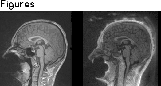
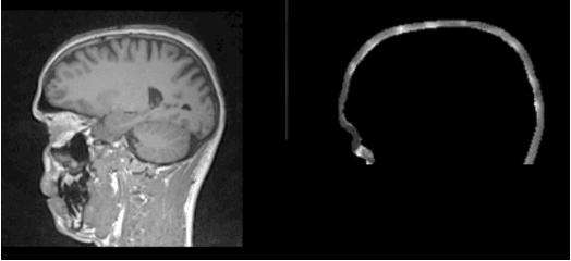
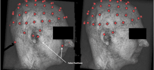
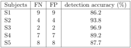
Simultaneous EEG/fMRI acquisition allows to measure brain activity at high spatial-temporal resolution. The localisation of EEG sources depends on several parameters including the position of the electrodes on the scalp. The position of the MR electrodes during its acquisitions is obtained with the use of the UTE sequence allowing their visualisation. The retrieval of the electrodes consists in obtaining the volume where the electrodes are located by applying a sphere detection algorithm. We detect around 90% of electrodes for each subject, and our UTE-based electrode detection showed an average position error of 3.7mm for all subjects.