Christian Barillot
VisAGeS
Effectiveness of regional diffusion MRI measures in distinguishing multiple sclerosis abnormalities within the cervical spinal cord
Aug 09, 2021


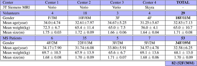
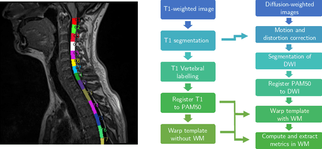
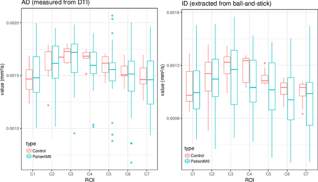
Abstract:Multiple sclerosis is an inflammatory disorder of the central nervous system. Quantitative MRI has huge potential to provide intrinsic and normative values of tissue properties useful for diagnosis, prognosis and ultimately clinical follow-up of this disease. However, there is a large discrepancy between the clinical observations and how the pathology is exhibited in MRI brain scans. Complementary to brain imaging, the study of multiple sclerosis lesions in the spinal cord has recently gained interest as a potential marker for early physical impairment. Therefore, investigating how the spinal cord is damaged using quantitative imaging, in particular, diffusion MRI, becomes an acute challenge. In this work, we extract average diffusion MRI metrics per vertebral level from spinal cord data acquired from multiple clinical sites. The diffusion-based metrics involved are extracted from the diffusion tensor imaging and Ball-and-Stick models and quantified for every cervical vertebral level using a collection of image processing methods and an atlas-based approach. Then, we perform a statistical analysis study to characterize the feasibility of these metrics to detect lesions. Specifically, we study the usefulness of combining different metrics to improve the accuracy prediction score associated with the presence of multiple sclerosis lesions. We demonstrate the grade of sensitivity to underlying microstructure changes in MS patients of each metric. Ball-and-Stick provides novel information about the MS damage to tissue microstructure. In addition, we show that choosing a subset of metrics: [FA, RD, MD] and [FWW, MD, Stick-AD, RD], which bring complementary information, has significantly increased the prediction score of the presence of the MS lesion in the cervical spinal cord.
Evaluation of distortion correction methods in diffusion MRI of the spinal cord
Aug 09, 2021


Abstract:Background: Magnetic field inhomogeneities generate important geometric distortions in reconstructed echo-planar images. Various procedures were proposed for correcting these distortions on brain images; yet, few neuroimaging studies tailored and incorporated the use of these techniques in spinal cord diffusion MRI. Purpose: We present a comparative evaluation of distortion correction methods that use the reversed gradient polarity technique on spinal cord. We propose novel geometric metrics to measure the alignment of the reconstructed diffusion model with the apparent centerline of the spinal cord. Subjects: 95 subjects, among which 29 healthy controls and 66 multiple sclerosis patients. Assessment: Geometric distortions were corrected using 4 state-of-the-art methods. We measured the alignment of the principal direction of diffusion with the apparent centerline of the spine after correction and the correlation with the reference anatomical image. Results are computed per vertebral level, to evaluate the impact on different portions of the spine. Besides, subjective evaluation of the quality of the correction of healthy subjects images was performed by three expert raters. Results: As a result of distortion correction, the diffusion directions are better aligned locally with the centerline, in particular at both ends of the acquisition window. The cross-correlation with anatomical image is also improved by Hyperelastic Susceptibility Artefact Correction (HySCO) and block-matching. The subjective evaluation for HySCO is significantly better (p < 0.05) than for Block-Matching; TOPUP performs significantly worse than the three other methods. Conclusion: Correction based on HySCO provide best results among the selected methods.
Reproducibility and Evolution of Diffusion MRI Measurements within the Cervical Spinal Cord in Multiple Sclerosis
Aug 08, 2021


Abstract:In Multiple Sclerosis (MS), there is a large discrepancy between the clinical observations and how the pathology is exhibited on brain images, this is known as the clinical-radiological paradox (CRP). One of the hypotheses is that the clinical deficit may be more related to the spinal cord damage than the number or location of lesions in the brain. Therefore, investigating how the spinal cord is damaged becomes an acute challenge to better understand and overcome the CRP. Diffusion MRI is known to provide quantitative figures of neuronal degeneration and axonal loss, in the brain as well as in the spinal cord. In this paper, we propose to investigate how diffusion MRI metrics vary in the different cervical regions with the progression of the disease. We first study the reproducibility of diffusion MRI on healthy volunteers with a test-retest procedure using both standard diffusion tensor imaging (DTI) and multi-compartment Ball-and-Stick models. Then, based on the test re-test quantitative calibration, we provide quantitative figures of pathology evolution between M0 and M12 in the cervical spine on a set of 31 MS patients, exhibiting how the pathology damage spans in the cervical spinal cord.
Automatic Electrodes Detection during simultaneous EEG/fMRI acquisition
Sep 17, 2018


Abstract:Simultaneous EEG/fMRI acquisition allows to measure brain activity at high spatial-temporal resolution. The localisation of EEG sources depends on several parameters including the position of the electrodes on the scalp. The position of the MR electrodes during its acquisitions is obtained with the use of the UTE sequence allowing their visualisation. The retrieval of the electrodes consists in obtaining the volume where the electrodes are located by applying a sphere detection algorithm. We detect around 90% of electrodes for each subject, and our UTE-based electrode detection showed an average position error of 3.7mm for all subjects.
 Add to Chrome
 Add to Firefox
 Add to Edge