Marcos Ortega
Weakly-supervised detection of AMD-related lesions in color fundus images using explainable deep learning
Dec 04, 2022Abstract:Age-related macular degeneration (AMD) is a degenerative disorder affecting the macula, a key area of the retina for visual acuity. Nowadays, it is the most frequent cause of blindness in developed countries. Although some promising treatments have been developed, their effectiveness is low in advanced stages. This emphasizes the importance of large-scale screening programs. Nevertheless, implementing such programs for AMD is usually unfeasible, since the population at risk is large and the diagnosis is challenging. All this motivates the development of automatic methods. In this sense, several works have achieved positive results for AMD diagnosis using convolutional neural networks (CNNs). However, none incorporates explainability mechanisms, which limits their use in clinical practice. In that regard, we propose an explainable deep learning approach for the diagnosis of AMD via the joint identification of its associated retinal lesions. In our proposal, a CNN is trained end-to-end for the joint task using image-level labels. The provided lesion information is of clinical interest, as it allows to assess the developmental stage of AMD. Additionally, the approach allows to explain the diagnosis from the identified lesions. This is possible thanks to the use of a CNN with a custom setting that links the lesions and the diagnosis. Furthermore, the proposed setting also allows to obtain coarse lesion segmentation maps in a weakly-supervised way, further improving the explainability. The training data for the approach can be obtained without much extra work by clinicians. The experiments conducted demonstrate that our approach can identify AMD and its associated lesions satisfactorily, while providing adequate coarse segmentation maps for most common lesions.
Improving AMD diagnosis by the simultaneous identification of associated retinal lesions
May 22, 2022Abstract:Age-related Macular Degeneration (AMD) is the predominant cause of blindness in developed countries, specially in elderly people. Moreover, its prevalence is increasing due to the global population ageing. In this scenario, early detection is crucial to avert later vision impairment. Nonetheless, implementing large-scale screening programmes is usually not viable, since the population at-risk is large and the analysis must be performed by expert clinicians. Also, the diagnosis of AMD is considered to be particularly difficult, as it is characterized by many different lesions that, in many cases, resemble those of other macular diseases. To overcome these issues, several works have proposed automatic methods for the detection of AMD in retinography images, the most widely used modality for the screening of the disease. Nowadays, most of these works use Convolutional Neural Networks (CNNs) for the binary classification of images into AMD and non-AMD classes. In this work, we propose a novel approach based on CNNs that simultaneously performs AMD diagnosis and the classification of its potential lesions. This latter secondary task has not yet been addressed in this domain, and provides complementary useful information that improves the diagnosis performance and helps understanding the decision. A CNN model is trained using retinography images with image-level labels for both AMD and lesion presence, which are relatively easy to obtain. The experiments conducted in several public datasets show that the proposed approach improves the detection of AMD, while achieving satisfactory results in the identification of most lesions.
Multi-stage transfer learning for lung segmentation using portable X-ray devices for patients with COVID-19
Oct 30, 2020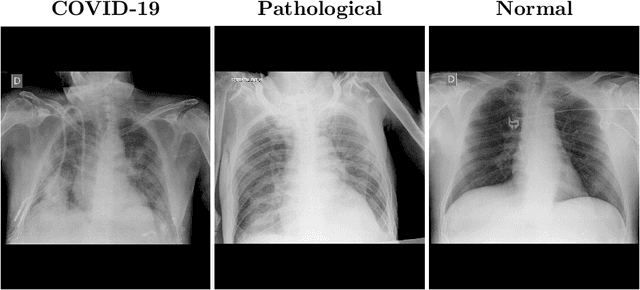
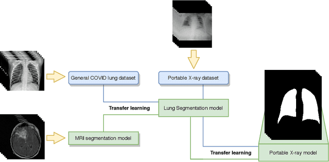

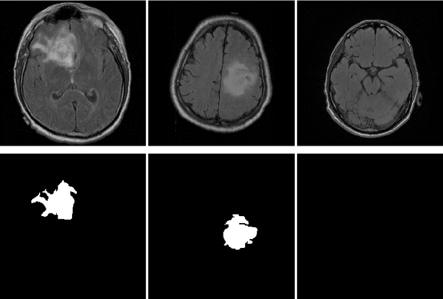
Abstract:In 2020, the SARS-CoV-2 virus causes a global pandemic of the new human coronavirus disease COVID-19. This pathogen primarily infects the respiratory system of the afflicted, usually resulting in pneumonia and in a severe case of acute respiratory distress syndrome. These disease developments result in the formation of different pathological structures in the lungs, similar to those observed in other viral pneumonias that can be detected by the use of chest X-rays. For this reason, the detection and analysis of the pulmonary regions, the main focus of affection of COVID-19, becomes a crucial part of both clinical and automatic diagnosis processes. Due to the overload of the health services, portable X-ray devices are widely used, representing an alternative to fixed devices to reduce the risk of cross-contamination. However, these devices entail different complications as the image quality that, together with the subjectivity of the clinician, make the diagnostic process more difficult. In this work, we developed a novel fully automatic methodology specially designed for the identification of these lung regions in X-ray images of low quality as those from portable devices. To do so, we took advantage of a large dataset from magnetic resonance imaging of a similar pathology and performed two stages of transfer learning to obtain a robust methodology with a low number of images from portable X-ray devices. This way, our methodology obtained a satisfactory accuracy of $0.9761 \pm 0.0100$ for patients with COVID-19, $0.9801 \pm 0.0104$ for normal patients and $0.9769 \pm 0.0111$ for patients with pulmonary diseases with similar characteristics as COVID-19 (such as pneumonia) but not genuine COVID-19.
Automatic segmentation of the Foveal Avascular Zone in ophthalmological OCT-A images
Nov 26, 2018


Abstract:Angiography by Optical Coherence Tomography is a non-invasive retinal imaging modality of recent appearance that allows the visualization of the vascular structure at predefined depths based on the detection of the blood movement. OCT-A images constitute a suitable scenario to analyse the retinal vascular properties of regions of interest, measuring the characteristics of the foveal vascular and avascular zones. Extracted parameters of this region can be used as prognostic factors that determine if the patient suffers from certain pathologies, indicating the associated pathological degree. The manual extraction of these biomedical parameters is a long, tedious and subjective process, introducing a significant intra and inter-expert variability, which penalizes the utility of the measurements. In addition, the absence of tools that automatically facilitate these calculations encourages the creation of computer-aided diagnosis frameworks that ease the doctor's work, increasing their productivity and making viable the use of this type of vascular biomarkers. We propose a fully automatic system that identifies and precisely segments the region of the foveal avascular zone (FAZ) using a novel ophthalmological image modality as is OCT-A. The system combines different image processing techniques to firstly identify the region where the FAZ is contained and, secondly, proceed with the extraction of its precise contour. The system was validated using a representative set of 168 OCT-A images, providing accurate results with the best correlation with the manual measurements of two experts clinician of 0.93 as well as a Jaccard's index of 0.82 of the best experimental case. This tool provides an accurate FAZ measurement with the desired objectivity and reproducibility, being very useful for the analysis of relevant vascular diseases through the study of the retinal microcirculation.
Multimodal Registration of Retinal Images Using Domain-Specific Landmarks and Vessel Enhancement
Apr 02, 2018

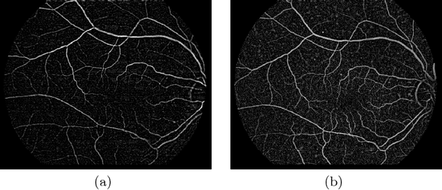
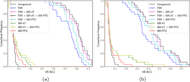
Abstract:The analysis of different image modalities is frequently performed in ophthalmology as it provides complementary information for the diagnosis and follow-up of relevant diseases, like hypertension or diabetes. This work presents a hybrid method for the multimodal registration of color fundus retinography and fluorescein angiography. The proposed method combines a feature-based approach, using domain-specific landmarks, with an intensity-based approach that employs a domain-adapted similarity metric. The methodology is tested on a dataset of 59 image pairs containing both healthy and pathological cases. The results show a satisfactory performance of the proposed combined approach in this multimodal scenario, improving the registration accuracy achieved by the feature-based and the intensity-based approaches.
 Add to Chrome
 Add to Firefox
 Add to Edge