Multi-stage transfer learning for lung segmentation using portable X-ray devices for patients with COVID-19
Paper and Code
Oct 30, 2020


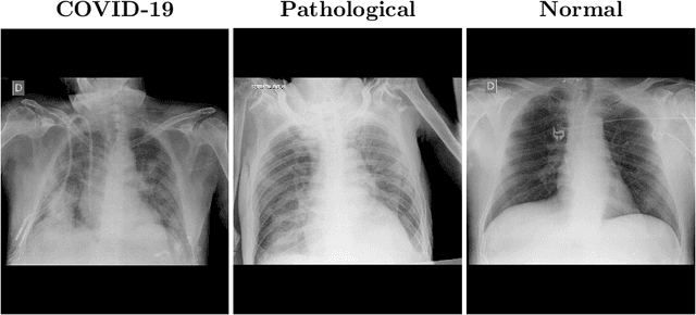
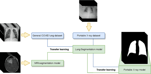
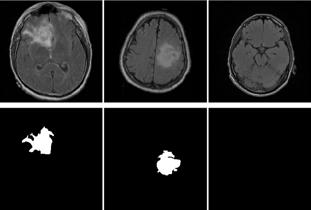
In 2020, the SARS-CoV-2 virus causes a global pandemic of the new human coronavirus disease COVID-19. This pathogen primarily infects the respiratory system of the afflicted, usually resulting in pneumonia and in a severe case of acute respiratory distress syndrome. These disease developments result in the formation of different pathological structures in the lungs, similar to those observed in other viral pneumonias that can be detected by the use of chest X-rays. For this reason, the detection and analysis of the pulmonary regions, the main focus of affection of COVID-19, becomes a crucial part of both clinical and automatic diagnosis processes. Due to the overload of the health services, portable X-ray devices are widely used, representing an alternative to fixed devices to reduce the risk of cross-contamination. However, these devices entail different complications as the image quality that, together with the subjectivity of the clinician, make the diagnostic process more difficult. In this work, we developed a novel fully automatic methodology specially designed for the identification of these lung regions in X-ray images of low quality as those from portable devices. To do so, we took advantage of a large dataset from magnetic resonance imaging of a similar pathology and performed two stages of transfer learning to obtain a robust methodology with a low number of images from portable X-ray devices. This way, our methodology obtained a satisfactory accuracy of $0.9761 \pm 0.0100$ for patients with COVID-19, $0.9801 \pm 0.0104$ for normal patients and $0.9769 \pm 0.0111$ for patients with pulmonary diseases with similar characteristics as COVID-19 (such as pneumonia) but not genuine COVID-19.
 Add to Chrome
 Add to Firefox
 Add to Edge