Klaus Scheffler
DISGAN: Wavelet-informed Discriminator Guides GAN to MRI Super-resolution with Noise Cleaning
Aug 23, 2023Abstract:MRI super-resolution (SR) and denoising tasks are fundamental challenges in the field of deep learning, which have traditionally been treated as distinct tasks with separate paired training data. In this paper, we propose an innovative method that addresses both tasks simultaneously using a single deep learning model, eliminating the need for explicitly paired noisy and clean images during training. Our proposed model is primarily trained for SR, but also exhibits remarkable noise-cleaning capabilities in the super-resolved images. Instead of conventional approaches that introduce frequency-related operations into the generative process, our novel approach involves the use of a GAN model guided by a frequency-informed discriminator. To achieve this, we harness the power of the 3D Discrete Wavelet Transform (DWT) operation as a frequency constraint within the GAN framework for the SR task on magnetic resonance imaging (MRI) data. Specifically, our contributions include: 1) a 3D generator based on residual-in-residual connected blocks; 2) the integration of the 3D DWT with $1\times 1$ convolution into a DWT+conv unit within a 3D Unet for the discriminator; 3) the use of the trained model for high-quality image SR, accompanied by an intrinsic denoising process. We dub the model "Denoising Induced Super-resolution GAN (DISGAN)" due to its dual effects of SR image generation and simultaneous denoising. Departing from the traditional approach of training SR and denoising tasks as separate models, our proposed DISGAN is trained only on the SR task, but also achieves exceptional performance in denoising. The model is trained on 3D MRI data from dozens of subjects from the Human Connectome Project (HCP) and further evaluated on previously unseen MRI data from subjects with brain tumours and epilepsy to assess its denoising and SR performance.
Pretraining is All You Need: A Multi-Atlas Enhanced Transformer Framework for Autism Spectrum Disorder Classification
Jul 04, 2023Abstract:Autism spectrum disorder (ASD) is a prevalent psychiatric condition characterized by atypical cognitive, emotional, and social patterns. Timely and accurate diagnosis is crucial for effective interventions and improved outcomes in individuals with ASD. In this study, we propose a novel Multi-Atlas Enhanced Transformer framework, METAFormer, ASD classification. Our framework utilizes resting-state functional magnetic resonance imaging data from the ABIDE I dataset, comprising 406 ASD and 476 typical control (TC) subjects. METAFormer employs a multi-atlas approach, where flattened connectivity matrices from the AAL, CC200, and DOS160 atlases serve as input to the transformer encoder. Notably, we demonstrate that self-supervised pretraining, involving the reconstruction of masked values from the input, significantly enhances classification performance without the need for additional or separate training data. Through stratified cross-validation, we evaluate the proposed framework and show that it surpasses state-of-the-art performance on the ABIDE I dataset, with an average accuracy of 83.7% and an AUC-score of 0.832. The code for our framework is available at https://github.com/Lugges991/METAFormer
A Three-Player GAN for Super-Resolution in Magnetic Resonance Imaging
Mar 24, 2023Abstract:Learning based single image super resolution (SISR) task is well investigated in 2D images. However, SISR for 3D Magnetics Resonance Images (MRI) is more challenging compared to 2D, mainly due to the increased number of neural network parameters, the larger memory requirement and the limited amount of available training data. Current SISR methods for 3D volumetric images are based on Generative Adversarial Networks (GANs), especially Wasserstein GANs due to their training stability. Other common architectures in the 2D domain, e.g. transformer models, require large amounts of training data and are therefore not suitable for the limited 3D data. However, Wasserstein GANs can be problematic because they may not converge to a global optimum and thus produce blurry results. Here, we propose a new method for 3D SR based on the GAN framework. Specifically, we use instance noise to balance the GAN training. Furthermore, we use a relativistic GAN loss function and an updating feature extractor during the training process. We show that our method produces highly accurate results. We also show that we need very few training samples. In particular, we need less than 30 samples instead of thousands of training samples that are typically required in previous studies. Finally, we show improved out-of-sample results produced by our model.
Prediction of motion induced magnetic fields for human brain MRI at 3T
Sep 30, 2022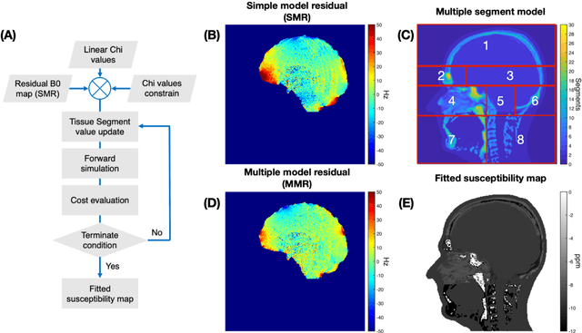
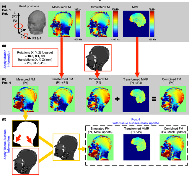
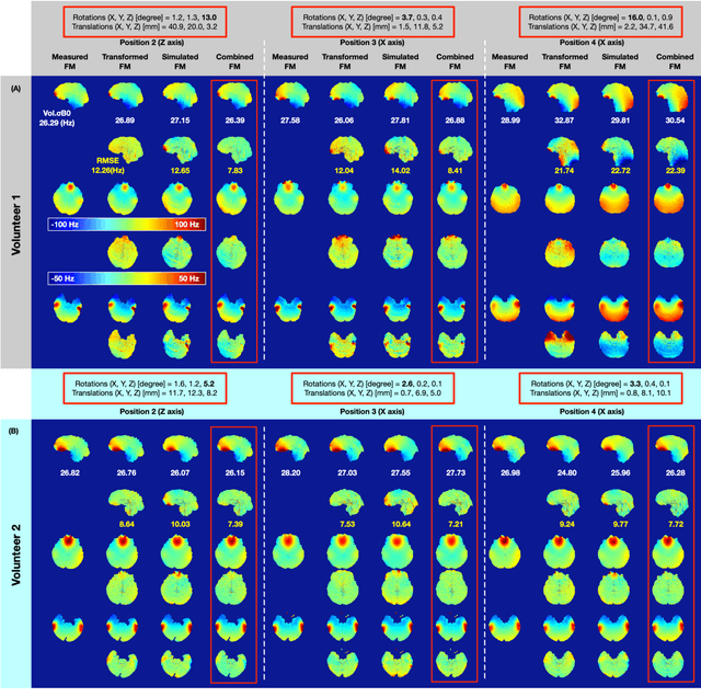
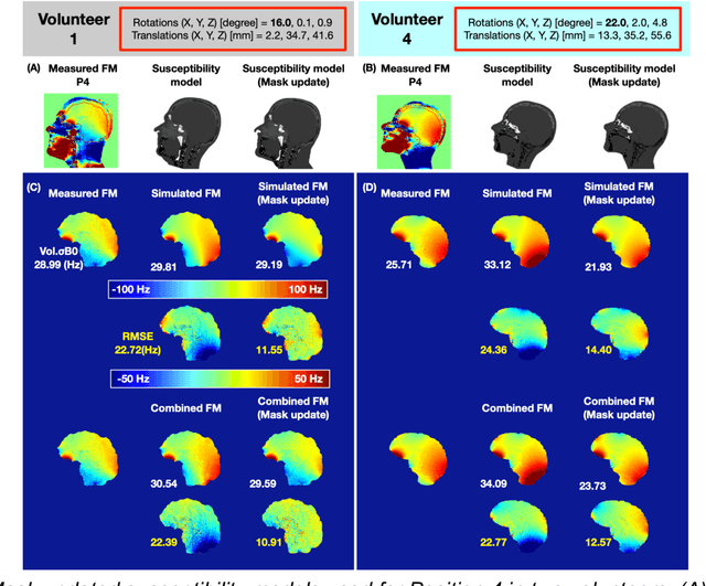
Abstract:Objective Maps of B0 field inhomogeneities are often used to improve MRI image quality, even in a retrospective fashion. These field inhomogeneities depend on the exact head position within the static field but acquiring field maps (FM) at every position is time consuming. Here we explore different ways to obtain B0 predictions at different head positions. Methods FM were predicted from iterative simulations with four field factors: 1) sample induced B0 field, 2) system's spherical harmonic shim field, 3) perturbing field originating outside the field of view, 4) sequence phase errors. The simulation was improved by including local susceptibility sources estimated from UTE scans and position-specific masks. The estimation performance of the simulated FMs and a transformed FM, obtained from the measured reference FM, were compared with the actual FM at different head positions. Results The transformed FM provided inconsistent results for large head movements (>5 degree rotation), while the simulation strategy had a superior prediction accuracy for all positions. The simulated FM was used to optimize B0 shims with up to 22.2% improvement with respect to the transformed FM approach. Conclusion The proposed simulation strategy is able to predict movement induced B0 field inhomogeneities yielding more precise estimates of the ground truth field homogeneity than the transformed FM.
Improving 3D convolutional neural network comprehensibility via interactive visualization of relevance maps: Evaluation in Alzheimer's disease
Dec 18, 2020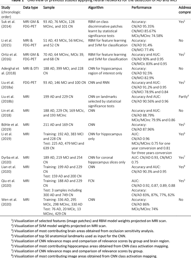

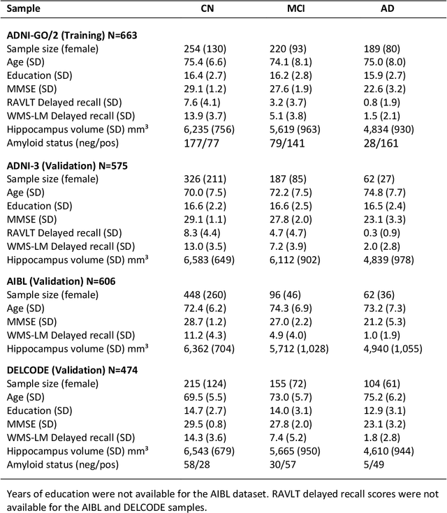
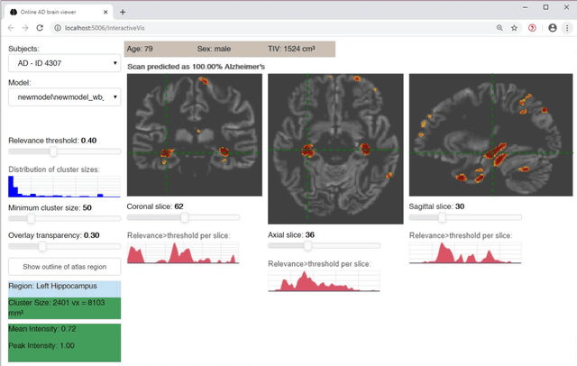
Abstract:Although convolutional neural networks (CNN) achieve high diagnostic accuracy for detecting Alzheimer's disease (AD) dementia based on magnetic resonance imaging (MRI) scans, they are not yet applied in clinical routine. One important reason for this is a lack of model comprehensibility. Recently developed visualization methods for deriving CNN relevance maps may help to fill this gap. We investigated whether models with higher accuracy also rely more on discriminative brain regions predefined by prior knowledge. We trained a CNN for the detection of AD in N=663 T1-weighted MRI scans of patients with dementia and amnestic mild cognitive impairment (MCI) and verified the accuracy of the models via cross-validation and in three independent samples including N=1655 cases. We evaluated the association of relevance scores and hippocampus volume to validate the clinical utility of this approach. To improve model comprehensibility, we implemented an interactive visualization of 3D CNN relevance maps. Across three independent datasets, group separation showed high accuracy for AD dementia vs. controls (AUC$\geq$0.92) and moderate accuracy for MCI vs. controls (AUC$\approx$0.75). Relevance maps indicated that hippocampal atrophy was considered as the most informative factor for AD detection, with additional contributions from atrophy in other cortical and subcortical regions. Relevance scores within the hippocampus were highly correlated with hippocampal volumes (Pearson's r$\approx$-0.81). The relevance maps highlighted atrophy in regions that we had hypothesized a priori. This strengthens the comprehensibility of the CNN models, which were trained in a purely data-driven manner based on the scans and diagnosis labels. The high hippocampus relevance scores and high performance achieved in independent samples support the validity of the CNN models in the detection of AD-related MRI abnormalities.
 Add to Chrome
 Add to Firefox
 Add to Edge