Jingfeng Lu
Ultrafast Cardiac Imaging Using Deep Learning For Speckle-Tracking Echocardiography
Jun 25, 2023
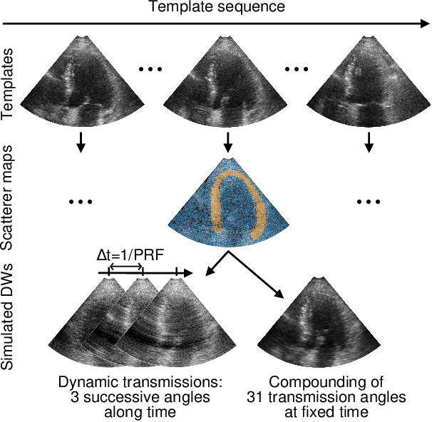
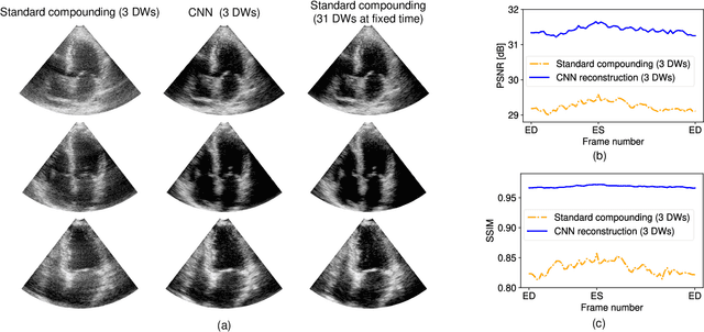
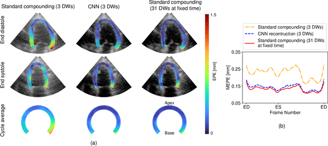
Abstract:High-quality ultrafast ultrasound imaging is based on coherent compounding from multiple transmissions of plane waves (PW) or diverging waves (DW). However, compounding results in reduced frame rate, as well as destructive interferences from high-velocity tissue motion if motion compensation (MoCo) is not considered. While many studies have recently shown the interest of deep learning for the reconstruction of high-quality static images from PW or DW, its ability to achieve such performance while maintaining the capability of tracking cardiac motion has yet to be assessed. In this paper, we addressed such issue by deploying a complex-weighted convolutional neural network (CNN) for image reconstruction and a state-of-the-art speckle tracking method. The evaluation of this approach was first performed by designing an adapted simulation framework, which provides specific reference data, i.e. high quality, motion artifact-free cardiac images. The obtained results showed that, while using only three DWs as input, the CNN-based approach yielded an image quality and a motion accuracy equivalent to those obtained by compounding 31 DWs free of motion artifacts. The performance was then further evaluated on non-simulated, experimental in vitro data, using a spinning disk phantom. This experiment demonstrated that our approach yielded high-quality image reconstruction and motion estimation, under a large range of velocities and outperforms a state-of-the-art MoCo-based approach at high velocities. Our method was finally assessed on in vivo datasets and showed consistent improvement in image quality and motion estimation compared to standard compounding. This demonstrates the feasibility and effectiveness of deep learning reconstruction for ultrafast speckle-tracking echocardiography.
High-quality Low-dose CT Reconstruction Using Convolutional Neural Networks with Spatial and Channel Squeeze and Excitation
Apr 01, 2021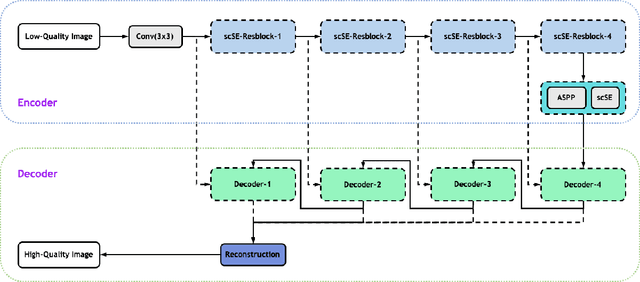
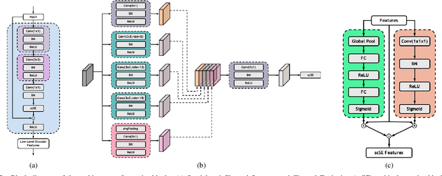
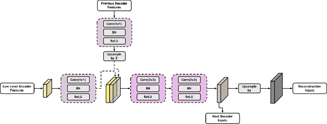
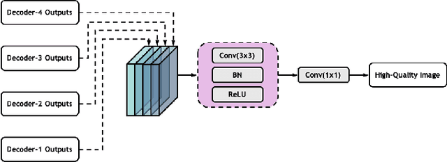
Abstract:Low-dose computed tomography (CT) allows the reduction of radiation risk in clinical applications at the expense of image quality, which deteriorates the diagnosis accuracy of radiologists. In this work, we present a High-Quality Imaging network (HQINet) for the CT image reconstruction from Low-dose computed tomography (CT) acquisitions. HQINet was a convolutional encoder-decoder architecture, where the encoder was used to extract spatial and temporal information from three contiguous slices while the decoder was used to recover the spacial information of the middle slice. We provide experimental results on the real projection data from low-dose CT Image and Projection Data (LDCT-and-Projection-data), demonstrating that the proposed approach yielded a notable improvement of the performance in terms of image quality, with a rise of 5.5dB in terms of peak signal-to-noise ratio (PSNR) and 0.29 in terms of mutual information (MI).
 Add to Chrome
 Add to Firefox
 Add to Edge