Jiayu Peng
Learning Ordering in Crystalline Materials with Symmetry-Aware Graph Neural Networks
Sep 20, 2024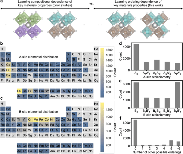
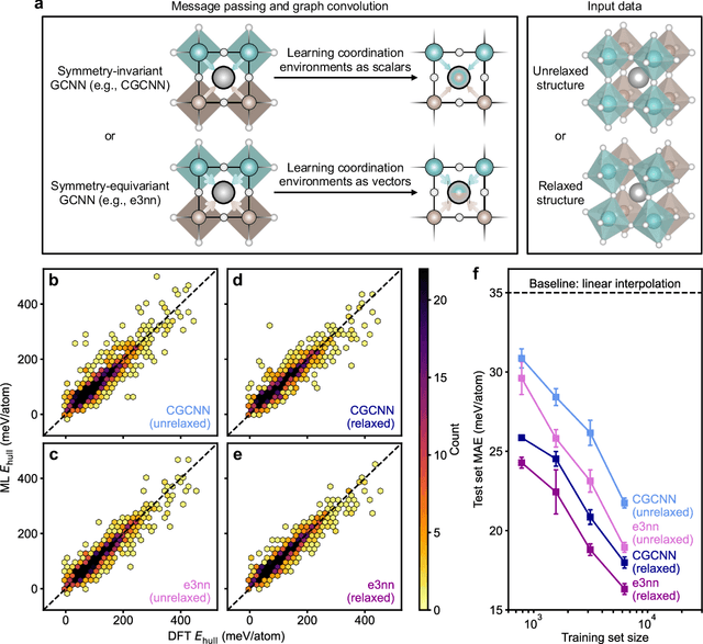
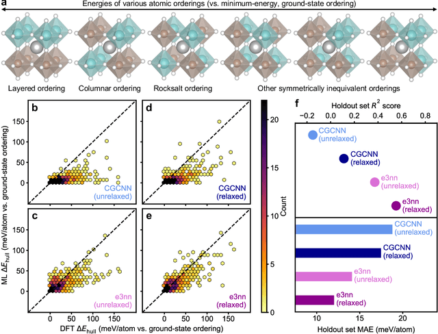
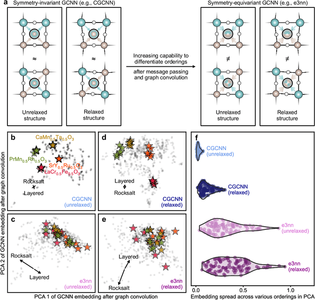
Abstract:Graph convolutional neural networks (GCNNs) have become a machine learning workhorse for screening the chemical space of crystalline materials in fields such as catalysis and energy storage, by predicting properties from structures. Multicomponent materials, however, present a unique challenge since they can exhibit chemical (dis)order, where a given lattice structure can encompass a variety of elemental arrangements ranging from highly ordered structures to fully disordered solid solutions. Critically, properties like stability, strength, and catalytic performance depend not only on structures but also on orderings. To enable rigorous materials design, it is thus critical to ensure GCNNs are capable of distinguishing among atomic orderings. However, the ordering-aware capability of GCNNs has been poorly understood. Here, we benchmark various neural network architectures for capturing the ordering-dependent energetics of multicomponent materials in a custom-made dataset generated with high-throughput atomistic simulations. Conventional symmetry-invariant GCNNs were found unable to discern the structural difference between the diverse symmetrically inequivalent atomic orderings of the same material, while symmetry-equivariant model architectures could inherently preserve and differentiate the distinct crystallographic symmetries of various orderings.
One-Stop Automated Diagnostic System for Carpal Tunnel Syndrome in Ultrasound Images Using Deep Learning
Feb 08, 2024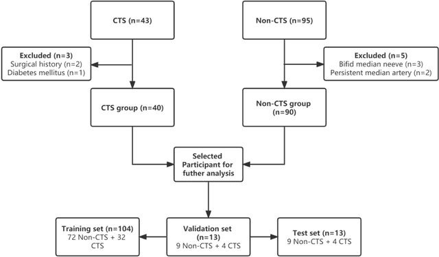
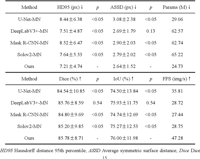

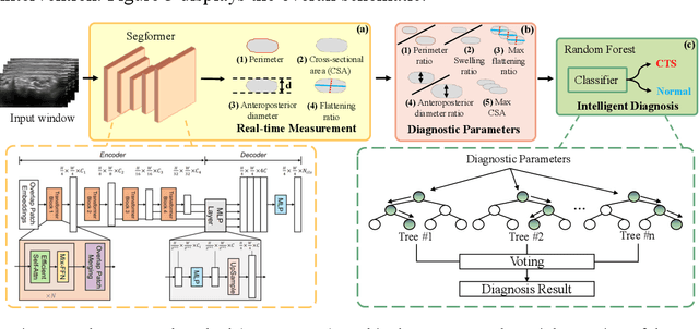
Abstract:Objective: Ultrasound (US) examination has unique advantages in diagnosing carpal tunnel syndrome (CTS) while identifying the median nerve (MN) and diagnosing CTS depends heavily on the expertise of examiners. To alleviate this problem, we aimed to develop a one-stop automated CTS diagnosis system (OSA-CTSD) and evaluate its effectiveness as a computer-aided diagnostic tool. Methods: We combined real-time MN delineation, accurate biometric measurements, and explainable CTS diagnosis into a unified framework, called OSA-CTSD. We collected a total of 32,301 static images from US videos of 90 normal wrists and 40 CTS wrists for evaluation using a simplified scanning protocol. Results: The proposed model showed better segmentation and measurement performance than competing methods, reporting that HD95 score of 7.21px, ASSD score of 2.64px, Dice score of 85.78%, and IoU score of 76.00%, respectively. In the reader study, it demonstrated comparable performance with the average performance of the experienced in classifying the CTS, while outperformed that of the inexperienced radiologists in terms of classification metrics (e.g., accuracy score of 3.59% higher and F1 score of 5.85% higher). Conclusion: The OSA-CTSD demonstrated promising diagnostic performance with the advantages of real-time, automation, and clinical interpretability. The application of such a tool can not only reduce reliance on the expertise of examiners, but also can help to promote the future standardization of the CTS diagnosis process, benefiting both patients and radiologists.
* Accepted by Ultrasound in Medicine & Biology
 Add to Chrome
 Add to Firefox
 Add to Edge