 Add to Chrome
 Add to Firefox
 Add to Edge
Reinforcement Learning for Ultrasound Image Analysis A Comprehensive Review of Advances and Applications
Feb 20, 2025Over the last decade, the use of machine learning (ML) approaches in medicinal applications has increased manifold. Most of these approaches are based on deep learning, which aims to learn representations from grid data (like medical images). However, reinforcement learning (RL) applications in medicine are relatively less explored. Medical applications often involve a sequence of subtasks that form a diagnostic pipeline, and RL is uniquely suited to optimize over such sequential decision-making tasks. Ultrasound (US) image analysis is a quintessential example of such a sequential decision-making task, where the raw signal captured by the US transducer undergoes a series of signal processing and image post-processing steps, generally leading to a diagnostic suggestion. The application of RL in US remains limited. Deep Reinforcement Learning (DRL), that combines deep learning and RL, holds great promise in optimizing these pipelines by enabling intelligent and sequential decision-making. This review paper surveys the applications of RL in US over the last decade. We provide a succinct overview of the theoretic framework of RL and its application in US image processing and review existing work in each aspect of the image analysis pipeline. A comprehensive search of Scopus filtered on relevance yielded 14 papers most relevant to this topic. These papers were further categorized based on their target applications image classification, image segmentation, image enhancement, video summarization, and auto navigation and path planning. We also examined the type of RL approach used in each publication. Finally, we discuss key areas in healthcare where DRL approaches in US could be used for sequential decision-making. We analyze the opportunities, challenges, and limitations, providing insights into the future potential of DRL in US image analysis.
Physics Driven Domain Specific Transporter Framework with Attention Mechanism for Ultrasound Imaging
Sep 13, 2021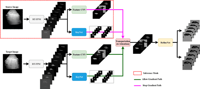
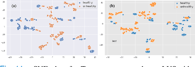
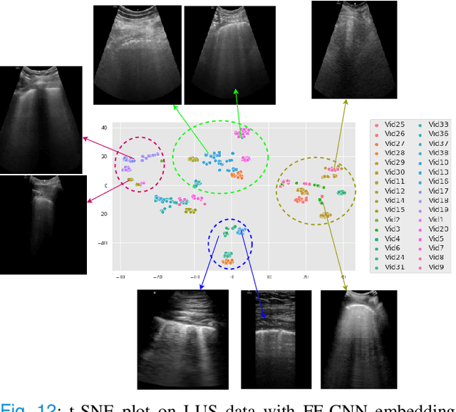
Most applications of deep learning techniques in medical imaging are supervised and require a large number of labeled data which is expensive and requires many hours of careful annotation by experts. In this paper, we propose an unsupervised, physics driven domain specific transporter framework with an attention mechanism to identify relevant key points with applications in ultrasound imaging. The proposed framework identifies key points that provide a concise geometric representation highlighting regions with high structural variation in ultrasound videos. We incorporate physics driven domain specific information as a feature probability map and use the radon transform to highlight features in specific orientations. The proposed framework has been trained on130 Lung ultrasound (LUS) videos and 113 Wrist ultrasound (WUS) videos and validated on 100 Lung ultrasound (LUS) videos and 58 Wrist ultrasound (WUS) videos acquired from multiple centers across the globe. Images from both datasets were independently assessed by experts to identify clinically relevant features such as A-lines, B-lines and pleura from LUS and radial metaphysis, radial epiphysis and carpal bones from WUS videos. The key points detected from both datasets showed high sensitivity (LUS = 99\% , WUS = 74\%) in detecting the image landmarks identified by experts. Also, on employing for classification of the given lung image into normal and abnormal classes, the proposed approach, even with no prior training, achieved an average accuracy of 97\% and an average F1-score of 95\% respectively on the task of co-classification with 3 fold cross-validation. With the purely unsupervised nature of the proposed approach, we expect the key point detection approach to increase the applicability of ultrasound in various examination performed in emergency and point of care.
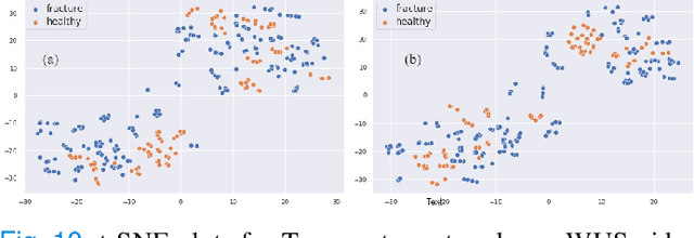
Domain Specific Transporter Framework to Detect Fractures in Ultrasound
Jun 09, 2021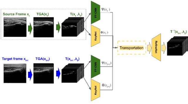
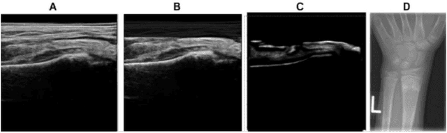
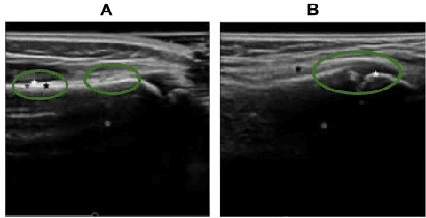
Ultrasound examination for detecting fractures is ideally suited for Emergency Departments (ED) as it is relatively fast, safe (from ionizing radiation), has dynamic imaging capability and is easily portable. High interobserver variability in manual assessment of ultrasound scans has piqued research interest in automatic assessment techniques using Deep Learning (DL). Most DL techniques are supervised and are trained on large numbers of labeled data which is expensive and requires many hours of careful annotation by experts. In this paper, we propose an unsupervised, domain specific transporter framework to identify relevant keypoints from wrist ultrasound scans. Our framework provides a concise geometric representation highlighting regions with high structural variation in a 3D ultrasound (3DUS) sequence. We also incorporate domain specific information represented by instantaneous local phase (LP) which detects bone features from 3DUS. We validate the technique on 3DUS videos obtained from 30 subjects. Each ultrasound scan was independently assessed by three readers to identify fractures along with the corresponding x-ray. Saliency of keypoints detected in the image\ are compared against manual assessment based on distance from relevant features.The transporter neural network was able to accurately detect 180 out of 250 bone regions sampled from wrist ultrasound videos. We expect this technique to increase the applicability of ultrasound in fracture detection.