Hossam El-Rewaidy
$\textbf{MyoMapNet}$: Accelerated Modified Look-Locker Inversion Recovery Myocardial T1 Mapping via Neural Networks
Mar 31, 2021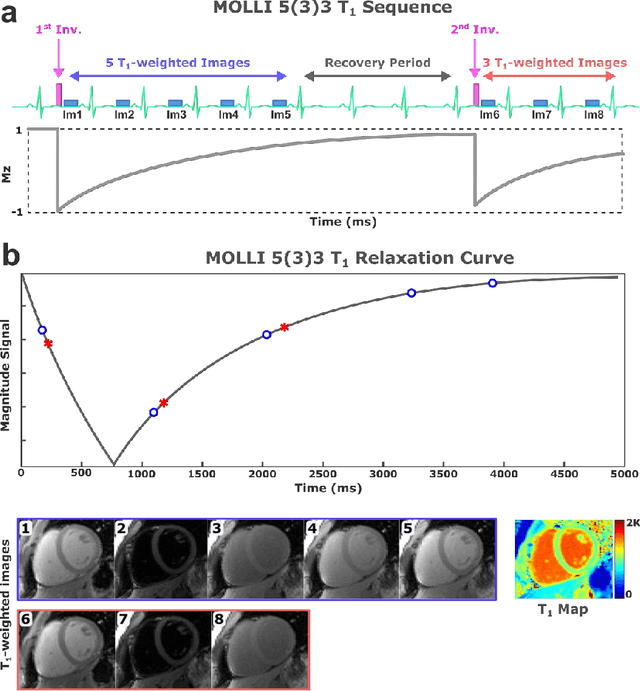
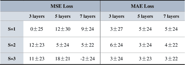
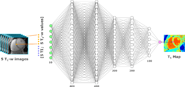

Abstract:Purpose: To develop and evaluate MyoMapNet, a rapid myocardial T1 mapping approach that uses neural networks (NN) to estimate voxel-wise myocardial T1 and extracellular (ECV) from T1-weighted images collected after a single inversion pulse over 4-5 heartbeats. Method: MyoMapNet utilizes a simple fully-connected NN to estimate T1 values from 5 (native) or 4 (post-contrast) T1-weighted images. Native MOLLI-5(3)3 T1 was collected in 717 subjects (386 males, 55$\pm$16.5 years) and post-contrast MOLLI-4(1)3(1)2 in 535 subjects (232 male, 56.5$\pm$15 years). The dataset was divided into training (80%) and testing (20%), where 20% of the training set was used to optimize MyoMapNet architecture (size and loss functions). We used MyoMapNet to estimate T1 and ECV maps with the first 5 (native) or 4 (post-contrast) T1-weighted images from the corresponding MOLLI sequence compared to the conventional and an abbreviated MOLLI using similar number of T1-weighted images with 3-parameter curve-fitting. Results: In our preliminary optimizaiton step, we determined that a 5-layers NN trained using mean-absolute-error loss yields lower estimation errors and was used subsequently in independent testing study. The myocardial T1 by MyoMapNet was similar to MOLLI (1200$\pm$45ms vs. 1199$\pm$46ms; P=0.3 for native T1, and 27.3$\pm$3.5% vs. 27.1$\pm$4%; P=0.4 for ECV). MyoMapNet had significantly smaller errors in T1 estimations compared to abbreviated-MOLLI (1$\pm$17ms vs. 31$\pm$34ms, P<0.01 for in native T1, and 0.1$\pm$1.3% vs. 1.9$\pm$2.5%, P<0.01 for ECV). The duration of T1 estimation was approximately 2 ms per slice using MyoMapNet. Conclusion: MyoMapNet T1 mapping enables myocardial T1 quantification in 4-5 heartbeats with near-instantaneous map estimation time with similar accuracy and precision as MOLLI. Keywords: Myocardial T1 mapping, MOLLI, T1 reconstruction, Neural network, Deep Learning.
Self-Supervised Physics-Guided Deep Learning Reconstruction For High-Resolution 3D LGE CMR
Nov 18, 2020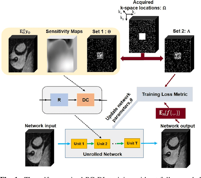
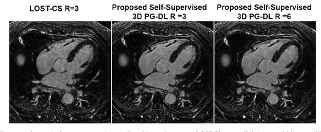
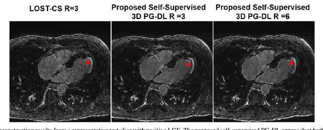
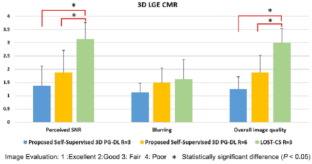
Abstract:Late gadolinium enhancement (LGE) cardiac MRI (CMR) is the clinical standard for diagnosis of myocardial scar. 3D isotropic LGE CMR provides improved coverage and resolution compared to 2D imaging. However, image acceleration is required due to long scan times and contrast washout. Physics-guided deep learning (PG-DL) approaches have recently emerged as an improved accelerated MRI strategy. Training of PG-DL methods is typically performed in supervised manner requiring fully-sampled data as reference, which is challenging in 3D LGE CMR. Recently, a self-supervised learning approach was proposed to enable training PG-DL techniques without fully-sampled data. In this work, we extend this self-supervised learning approach to 3D imaging, while tackling challenges related to small training database sizes of 3D volumes. Results and a reader study on prospectively accelerated 3D LGE show that the proposed approach at 6-fold acceleration outperforms the clinically utilized compressed sensing approach at 3-fold acceleration.
 Add to Chrome
 Add to Firefox
 Add to Edge