 Add to Chrome
 Add to Firefox
 Add to Edge
SynthRAR: Ring Artifacts Reduction in CT with Unrolled Network and Synthetic Data Training
Feb 12, 2026Defective and inconsistent responses in CT detectors can cause ring and streak artifacts in the reconstructed images, making them unusable for clinical purposes. In recent years, several ring artifact reduction solutions have been proposed in the image domain or in the sinogram domain using supervised deep learning methods. However, these methods require dedicated datasets for training, leading to a high data collection cost. Furthermore, existing approaches focus exclusively on either image-space or sinogram-space correction, neglecting the intrinsic correlations from the forward operation of the CT geometry. Based on the theoretical analysis of non-ideal CT detector responses, the RAR problem is reformulated as an inverse problem by using an unrolled network, which considers non-ideal response together with linear forward-projection with CT geometry. Additionally, the intrinsic correlations of ring artifacts between the sinogram and image domains are leveraged through synthetic data derived from natural images, enabling the trained model to correct artifacts without requiring real-world clinical data. Extensive evaluations on diverse scanning geometries and anatomical regions demonstrate that the model trained on synthetic data consistently outperforms existing state-of-the-art methods.
Text controllable PET denoising
Jan 28, 2026Positron Emission Tomography (PET) imaging is a vital tool in medical diagnostics, offering detailed insights into molecular processes within the human body. However, PET images often suffer from complicated noise, which can obscure critical diagnostic information. The quality of the PET image is impacted by various factors including scanner hardware, image reconstruction, tracer properties, dose/count level, and acquisition time. In this study, we propose a novel text-guided denoising method capable of enhancing PET images across a wide range of count levels within a single model. The model utilized the features from a pretrained CLIP model with a U-Net based denoising model. Experimental results demonstrate that the proposed model leads significant improvements in both qualitative and quantitative assessments. The flexibility of the model shows the potential for helping more complicated denoising demands or reducing the acquisition time.
Low performing pixel correction in computed tomography with unrolled network and synthetic data training
Jan 28, 2026Low performance pixels (LPP) in Computed Tomography (CT) detectors would lead to ring and streak artifacts in the reconstructed images, making them clinically unusable. In recent years, several solutions have been proposed to correct LPP artifacts, either in the image domain or in the sinogram domain using supervised deep learning methods. However, these methods require dedicated datasets for training, which are expensive to collect. Moreover, existing approaches focus solely either on image-space or sinogram-space correction, ignoring the intrinsic correlations from the forward operation of the CT geometry. In this work, we propose an unrolled dual-domain method based on synthetic data to correct LPP artifacts. Specifically, the intrinsic correlations of LPP between the sinogram and image domains are leveraged through synthetic data generated from natural images, enabling the trained model to correct artifacts without requiring any real-world clinical data. In experiments simulating 1-2% detectors defect near the isocenter, the proposed method outperformed the state-of-the-art approaches by a large margin. The results indicate that our solution can correct LPP artifacts without the cost of data collection for model training, and it is adaptable to different scanner settings for software-based applications.
SynBT: High-quality Tumor Synthesis for Breast Tumor Segmentation by 3D Diffusion Model
Sep 03, 2025Synthetic tumors in medical images offer controllable characteristics that facilitate the training of machine learning models, leading to an improved segmentation performance. However, the existing methods of tumor synthesis yield suboptimal performances when tumor occupies a large spatial volume, such as breast tumor segmentation in MRI with a large field-of-view (FOV), while commonly used tumor generation methods are based on small patches. In this paper, we propose a 3D medical diffusion model, called SynBT, to generate high-quality breast tumor (BT) in contrast-enhanced MRI images. The proposed model consists of a patch-to-volume autoencoder, which is able to compress the high-resolution MRIs into compact latent space, while preserving the resolution of volumes with large FOV. Using the obtained latent space feature vector, a mask-conditioned diffusion model is used to synthesize breast tumors within selected regions of breast tissue, resulting in realistic tumor appearances. We evaluated the proposed method for a tumor segmentation task, which demonstrated the proposed high-quality tumor synthesis method can facilitate the common segmentation models with performance improvement of 2-3% Dice Score on a large public dataset, and therefore provides benefits for tumor segmentation in MRI images.
Zero-shot Bias Correction: Efficient MR Image Inhomogeneity Reduction Without Any Data
Jan 21, 2025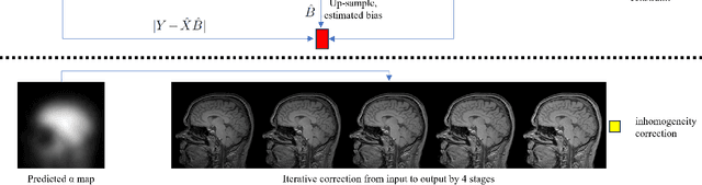


In recent years, deep neural networks for image inhomogeneity reduction have shown promising results. However, current methods with (un)supervised solutions require preparing a training dataset, which is expensive and laborious for data collection. In this work, we demonstrate a novel zero-shot deep neural networks, which requires no data for pre-training and dedicated assumption of the bias field. The designed light-weight CNN enables an efficient zero-shot adaptation for bias-corrupted image correction. Our method provides a novel solution to mitigate the biased corrupted image as iterative homogeneity refinement, which therefore ensures the considered issue can be solved easier with stable convergence of zero-shot optimization. Extensive comparison on different datasets show that the proposed method performs better than current data-free N4 methods in both efficiency and accuracy.
Quality Enhancement of Radiographic X-ray Images by Interpretable Mapping
Jan 21, 2025X-ray imaging is the most widely used medical imaging modality. However, in the common practice, inconsistency in the initial presentation of X-ray images is a common complaint by radiologists. Different patient positions, patient habitus and scanning protocols can lead to differences in image presentations, e.g., differences in brightness and contrast globally or regionally. To compensate for this, additional work will be executed by clinical experts to adjust the images to the desired presentation, which can be time-consuming. Existing deep-learning-based end-to-end solutions can automatically correct images with promising performances. Nevertheless, these methods are hard to be interpreted and difficult to be understood by clinical experts. In this manuscript, a novel interpretable mapping method by deep learning is proposed, which automatically enhances the image brightness and contrast globally and locally. Meanwhile, because the model is inspired by the workflow of the brightness and contrast manipulation, it can provide interpretable pixel maps for explaining the motivation of image enhancement. The experiment on the clinical datasets show the proposed method can provide consistent brightness and contrast correction on X-ray images with accuracy of 24.75 dB PSNR and 0.8431 SSIM.
Medical Instrument Segmentation in 3D US by Hybrid Constrained Semi-Supervised Learning
Jul 30, 2021


Medical instrument segmentation in 3D ultrasound is essential for image-guided intervention. However, to train a successful deep neural network for instrument segmentation, a large number of labeled images are required, which is expensive and time-consuming to obtain. In this article, we propose a semi-supervised learning (SSL) framework for instrument segmentation in 3D US, which requires much less annotation effort than the existing methods. To achieve the SSL learning, a Dual-UNet is proposed to segment the instrument. The Dual-UNet leverages unlabeled data using a novel hybrid loss function, consisting of uncertainty and contextual constraints. Specifically, the uncertainty constraints leverage the uncertainty estimation of the predictions of the UNet, and therefore improve the unlabeled information for SSL training. In addition, contextual constraints exploit the contextual information of the training images, which are used as the complementary information for voxel-wise uncertainty estimation. Extensive experiments on multiple ex-vivo and in-vivo datasets show that our proposed method achieves Dice score of about 68.6%-69.1% and the inference time of about 1 sec. per volume. These results are better than the state-of-the-art SSL methods and the inference time is comparable to the supervised approaches.
Weakly-supervised Learning For Catheter Segmentation in 3D Frustum Ultrasound
Oct 19, 2020


Accurate and efficient catheter segmentation in 3D ultrasound (US) is essential for cardiac intervention. Currently, the state-of-the-art segmentation algorithms are based on convolutional neural networks (CNNs), which achieved remarkable performances in a standard Cartesian volumetric data. Nevertheless, these approaches suffer the challenges of low efficiency and GPU unfriendly image size. Therefore, such difficulties and expensive hardware requirements become a bottleneck to build accurate and efficient segmentation models for real clinical application. In this paper, we propose a novel Frustum ultrasound based catheter segmentation method. Specifically, Frustum ultrasound is a polar coordinate based image, which includes same information of standard Cartesian image but has much smaller size, which overcomes the bottleneck of efficiency than conventional Cartesian images. Nevertheless, the irregular and deformed Frustum images lead to more efforts for accurate voxel-level annotation. To address this limitation, a weakly supervised learning framework is proposed, which only needs 3D bounding box annotations overlaying the region-of-interest to training the CNNs. Although the bounding box annotation includes noise and inaccurate annotation to mislead to model, it is addressed by the proposed pseudo label generated scheme. The labels of training voxels are generated by incorporating class activation maps with line filtering, which is iteratively updated during the training. Our experimental results show the proposed method achieved the state-of-the-art performance with an efficiency of 0.25 second per volume. More crucially, the Frustum image segmentation provides a much faster and cheaper solution for segmentation in 3D US image, which meet the demands of clinical applications.
Medical Instrument Detection in Ultrasound-Guided Interventions: A Review
Jul 09, 2020


Medical instrument detection is essential for computer-assisted interventions since it would facilitate the surgeons to find the instrument efficiently with a better interpretation, which leads to a better outcome. This article reviews medical instrument detection methods in the ultrasound-guided intervention. First, we present a comprehensive review of instrument detection methodologies, which include traditional non-data-driven methods and data-driven methods. The non-data-driven methods were extensively studied prior to the era of machine learning, i.e. data-driven approaches. We discuss the main clinical applications of medical instrument detection in ultrasound, including anesthesia, biopsy, prostate brachytherapy, and cardiac catheterization, which were validated on clinical datasets. Finally, we selected several principal publications to summarize the key issues and potential research directions for the computer-assisted intervention community.
Deep Q-Network-Driven Catheter Segmentation in 3D US by Hybrid Constrained Semi-Supervised Learning and Dual-UNet
Jun 25, 2020


Catheter segmentation in 3D ultrasound is important for computer-assisted cardiac intervention. However, a large amount of labeled images are required to train a successful deep convolutional neural network (CNN) to segment the catheter, which is expensive and time-consuming. In this paper, we propose a novel catheter segmentation approach, which requests fewer annotations than the supervised learning method, but nevertheless achieves better performance. Our scheme considers a deep Q learning as the pre-localization step, which avoids voxel-level annotation and which can efficiently localize the target catheter. With the detected catheter, patch-based Dual-UNet is applied to segment the catheter in 3D volumetric data. To train the Dual-UNet with limited labeled images and leverage information of unlabeled images, we propose a novel semi-supervised scheme, which exploits unlabeled images based on hybrid constraints from predictions. Experiments show the proposed scheme achieves a higher performance than state-of-the-art semi-supervised methods, while it demonstrates that our method is able to learn from large-scale unlabeled images.