Fangliang Bai
LSHR-Net: a hardware-friendly solution for high-resolution computational imaging using a mixed-weights neural network
Apr 27, 2020


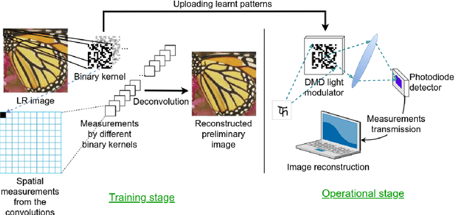
Abstract:Recent work showed neural-network-based approaches to reconstructing images from compressively sensed measurements offer significant improvements in accuracy and signal compression. Such methods can dramatically boost the capability of computational imaging hardware. However, to date, there have been two major drawbacks: (1) the high-precision real-valued sensing patterns proposed in the majority of existing works can prove problematic when used with computational imaging hardware such as a digital micromirror sampling device and (2) the network structures for image reconstruction involve intensive computation, which is also not suitable for hardware deployment. To address these problems, we propose a novel hardware-friendly solution based on mixed-weights neural networks for computational imaging. In particular, learned binary-weight sensing patterns are tailored to the sampling device. Moreover, we proposed a recursive network structure for low-resolution image sampling and high-resolution reconstruction scheme. It reduces both the required number of measurements and reconstruction computation by operating convolution on small intermediate feature maps. The recursive structure further reduced the model size, making the network more computationally efficient when deployed with the hardware. Our method has been validated on benchmark datasets and achieved the state of the art reconstruction accuracy. We tested our proposed network in conjunction with a proof-of-concept hardware setup.
Cystoid macular edema segmentation of Optical Coherence Tomography images using fully convolutional neural networks and fully connected CRFs
Sep 15, 2017


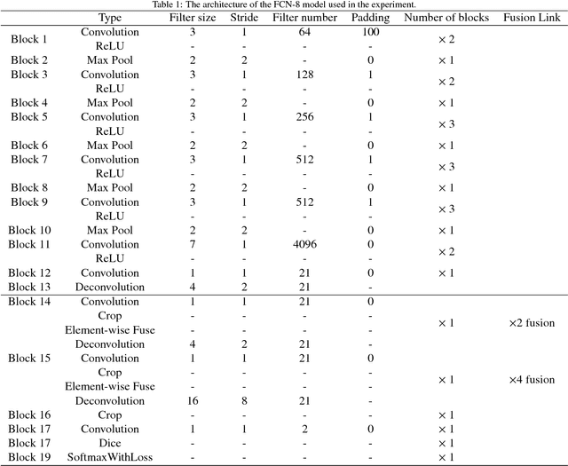
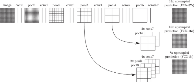
Abstract:In this paper we present a new method for cystoid macular edema (CME) segmentation in retinal Optical Coherence Tomography (OCT) images, using a fully convolutional neural network (FCN) and a fully connected conditional random fields (dense CRFs). As a first step, the framework trains the FCN model to extract features from retinal layers in OCT images, which exhibit CME, and then segments CME regions using the trained model. Thereafter, dense CRFs are used to refine the segmentation according to the edema appearance. We have trained and tested the framework with OCT images from 10 patients with diabetic macular edema (DME). Our experimental results show that fluid and concrete macular edema areas were segmented with good adherence to boundaries. A segmentation accuracy of $0.61\pm 0.21$ (Dice coefficient) was achieved, with respect to the ground truth, which compares favourably with the previous state-of-the-art that used a kernel regression based method ($0.51\pm 0.34$). Our approach is versatile and we believe it can be easily adapted to detect other macular defects.
 Add to Chrome
 Add to Firefox
 Add to Edge