Ethar Alzaid
Synergy vs. Noise: Performance-Guided Multimodal Fusion For Biochemical Recurrence-Free Survival in Prostate Cancer
Nov 14, 2025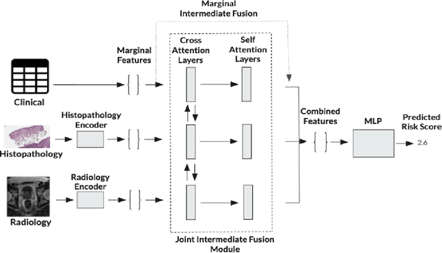
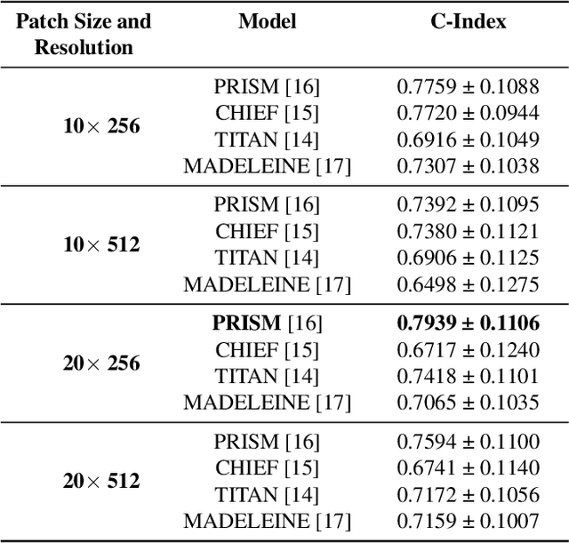
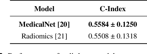
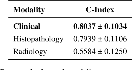
Abstract:Multimodal deep learning (MDL) has emerged as a transformative approach in computational pathology. By integrating complementary information from multiple data sources, MDL models have demonstrated superior predictive performance across diverse clinical tasks compared to unimodal models. However, the assumption that combining modalities inherently improves performance remains largely unexamined. We hypothesise that multimodal gains depend critically on the predictive quality of individual modalities, and that integrating weak modalities may introduce noise rather than complementary information. We test this hypothesis on a prostate cancer dataset with histopathology, radiology, and clinical data to predict time-to-biochemical recurrence. Our results confirm that combining high-performing modalities yield superior performance compared to unimodal approaches. However, integrating a poor-performing modality with other higher-performing modalities degrades predictive accuracy. These findings demonstrate that multimodal benefit requires selective, performance-guided integration rather than indiscriminate modality combination, with implications for MDL design across computational pathology and medical imaging.
MultiSurv: A Multimodal Deep Survival Framework for Prostrate and Bladder Cancer
Sep 05, 2025Abstract:Accurate prediction of time-to-event outcomes is a central challenge in oncology, with significant implications for treatment planning and patient management. In this work, we present MultiSurv, a multimodal deep survival model utilising DeepHit with a projection layer and inter-modality cross-attention, which integrates heterogeneous patient data, including clinical, MRI, RNA-seq and whole-slide pathology features. The model is designed to capture complementary prognostic signals across modalities and estimate individualised time-to-biochemical recurrence in prostate cancer and time-to-cancer recurrence in bladder cancer. Our approach was evaluated in the context of the CHIMERA Grand Challenge, across two of the three provided tasks. For Task 1 (prostate cancer bio-chemical recurrence prediction), the proposed framework achieved a concordance index (C-index) of 0.843 on 5-folds cross-validation and 0.818 on CHIMERA development set, demonstrating robust discriminatory ability. For Task 3 (bladder cancer recurrence prediction), the model obtained a C-index of 0.662 on 5-folds cross-validation and 0.457 on development set, highlighting its adaptability and potential for clinical translation. These results suggest that leveraging multimodal integration with deep survival learning provides a promising pathway toward personalised risk stratification in prostate and bladder cancer. Beyond the challenge setting, our framework is broadly applicable to survival prediction tasks involving heterogeneous biomedical data.
Large Multimodal Model based Standardisation of Pathology Reports with Confidence and their Prognostic Significance
May 03, 2024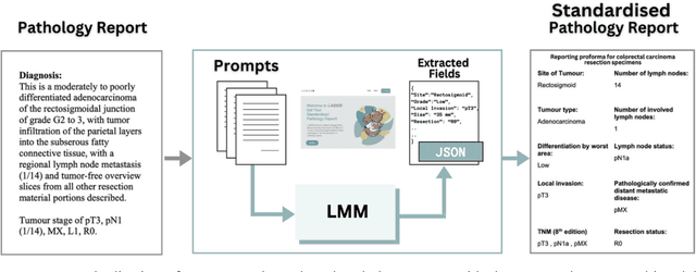

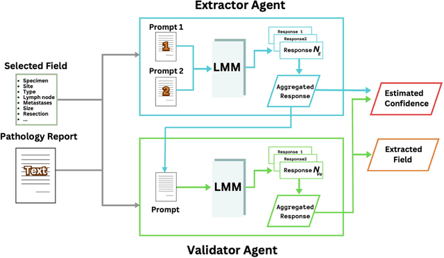
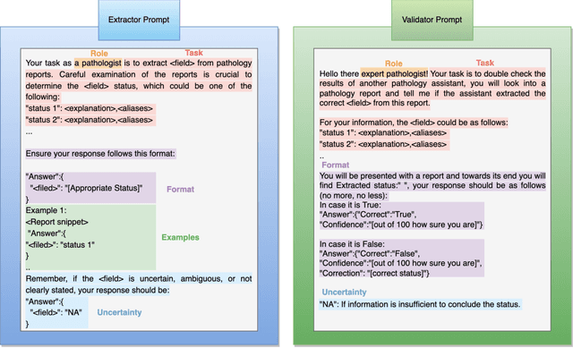
Abstract:Pathology reports are rich in clinical and pathological details but are often presented in free-text format. The unstructured nature of these reports presents a significant challenge limiting the accessibility of their content. In this work, we present a practical approach based on the use of large multimodal models (LMMs) for automatically extracting information from scanned images of pathology reports with the goal of generating a standardised report specifying the value of different fields along with estimated confidence about the accuracy of the extracted fields. The proposed approach overcomes limitations of existing methods which do not assign confidence scores to extracted fields limiting their practical use. The proposed framework uses two stages of prompting a Large Multimodal Model (LMM) for information extraction and validation. The framework generalises to textual reports from multiple medical centres as well as scanned images of legacy pathology reports. We show that the estimated confidence is an effective indicator of the accuracy of the extracted information that can be used to select only accurately extracted fields. We also show the prognostic significance of structured and unstructured data from pathology reports and show that the automatically extracted field values significant prognostic value for patient stratification. The framework is available for evaluation via the URL: https://labieb.dcs.warwick.ac.uk/.
TIAViz: A Browser-based Visualization Tool for Computational Pathology Models
Feb 15, 2024


Abstract:Digital pathology has gained significant traction in modern healthcare systems. This shift from optical microscopes to digital imagery brings with it the potential for improved diagnosis, efficiency, and the integration of AI tools into the pathologists workflow. A critical aspect of this is visualization. Throughout the development of a machine learning (ML) model in digital pathology, it is crucial to have flexible, openly available tools to visualize models, from their outputs and predictions to the underlying annotations and images used to train or test a model. We introduce TIAViz, a Python-based visualization tool built into TIAToolbox which allows flexible, interactive, fully zoomable overlay of a wide variety of information onto whole slide images, including graphs, heatmaps, segmentations, annotations and other WSIs. The UI is browser-based, allowing use either locally, on a remote machine, or on a server to provide publicly available demos. This tool is open source and is made available at: https://github.com/TissueImageAnalytics/tiatoolbox and via pip installation (pip install tiatoolbox) and conda as part of TIAToolbox.
 Add to Chrome
 Add to Firefox
 Add to Edge