Eldad Elnekave
Improved ICH classification using task-dependent learning
Jun 29, 2019


Abstract:Head CT is one of the most commonly performed imaging studied in the Emergency Department setting and Intracranial hemorrhage (ICH) is among the most critical and timesensitive findings to be detected on Head CT. We present BloodNet, a deep learning architecture designed for optimal triaging of Head CTs, with the goal of decreasing the time from CT acquisition to accurate ICH detection. The BloodNet architecture incorporates dependency between the otherwise independent tasks of segmentation and classification, achieving improved classification results. AUCs of 0.9493 and 0.9566 are reported on held out positive-enriched and randomly sampled sets comprised of over 1400 studies acquired from over 10 different hospitals. These results are comparable to previously reported results with smaller number of tagged studies.
PHT-bot: Deep-Learning based system for automatic risk stratification of COPD patients based upon signs of Pulmonary Hypertension
May 28, 2019Abstract:Chronic Obstructive Pulmonary Disease (COPD) is a leading cause of morbidity and mortality worldwide. Identifying those at highest risk of deterioration would allow more effective distribution of preventative and surveillance resources. Secondary pulmonary hypertension is a manifestation of advanced COPD, which can be reliably diagnosed by the main Pulmonary Artery (PA) to Ascending Aorta (Ao) ratio. In effect, a PA diameter to Ao diameter ratio of greater than 1 has been demonstrated to be a reliable marker of increased pulmonary arterial pressure. Although clinically valuable and readily visualized, the manual assessment of the PA and the Ao diameters is time consuming and under-reported. The present study describes a non invasive method to measure the diameters of both the Ao and the PA from contrast-enhanced chest Computed Tomography (CT). The solution applies deep learning techniques in order to select the correct axial slice to measure, and to segment both arteries. The system achieves test Pearson correlation coefficient scores of 93% for the Ao and 92% for the PA. To the best of our knowledge, it is the first such fully automated solution.
TextRay: Mining Clinical Reports to Gain a Broad Understanding of Chest X-rays
Jun 06, 2018


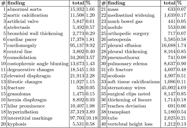
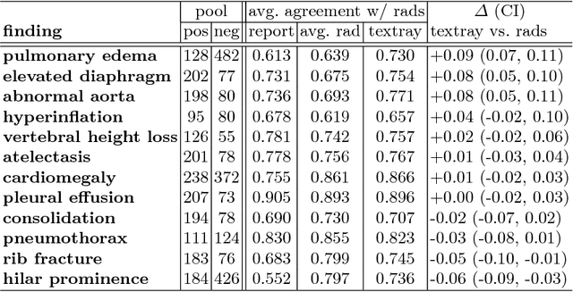
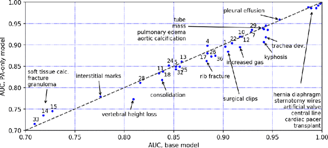
Abstract:The chest X-ray (CXR) is by far the most commonly performed radiological examination for screening and diagnosis of many cardiac and pulmonary diseases. There is an immense world-wide shortage of physicians capable of providing rapid and accurate interpretation of this study. A radiologist-driven analysis of over two million CXR reports generated an ontology including the 40 most prevalent pathologies on CXR. By manually tagging a relatively small set of sentences, we were able to construct a training set of 959k studies. A deep learning model was trained to predict the findings given the patient frontal and lateral scans. For 12 of the findings we compare the model performance against a team of radiologists and show that in most cases the radiologists agree on average more with the algorithm than with each other.
Compression Fractures Detection on CT
Jun 06, 2017Abstract:The presence of a vertebral compression fracture is highly indicative of osteoporosis and represents the single most robust predictor for development of a second osteoporotic fracture in the spine or elsewhere. Less than one third of vertebral compression fractures are diagnosed clinically. We present an automated method for detecting spine compression fractures in Computed Tomography (CT) scans. The algorithm is composed of three processes. First, the spinal column is segmented and sagittal patches are extracted. The patches are then binary classified using a Convolutional Neural Network (CNN). Finally a Recurrent Neural Network (RNN) is utilized to predict whether a vertebral fracture is present in the series of patches.
 Add to Chrome
 Add to Firefox
 Add to Edge