Ebrahim Nasr-Esfahani
Dense Fully Convolutional Network for Skin Lesion Segmentation
Jun 05, 2018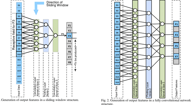

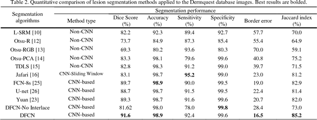
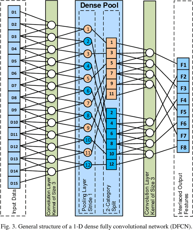
Abstract:Lesion segmentation in skin images is an important step in computerized detection of skin cancer. Melanoma is known as one of the most life threatening types of this cancer. Existing methods often fall short of accurately segmenting lesions with fuzzy boarders. In this paper, a new class of fully convolutional network is proposed, with new dense pooling layers for segmentation of lesion regions in non-dermoscopic images. Unlike other existing convolutional networks, this proposed network is designed to produce dense feature maps. This network leads to highly accurate segmentation of lesions. The produced dice score here is 91.6% which outperforms state-of-the-art algorithms in segmentation of skin lesions based on the Dermquest dataset.
Liver segmentation in CT images using three dimensional to two dimensional fully convolutional network
Mar 03, 2018
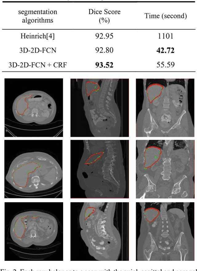
Abstract:The need for CT scan analysis is growing for pre-diagnosis and therapy of abdominal organs. Automatic organ segmentation of abdominal CT scan can help radiologists analyze the scans faster and segment organ images with fewer errors. However, existing methods are not efficient enough to perform the segmentation process for victims of accidents and emergencies situations. In this paper we propose an efficient liver segmentation with our 3D to 2D fully connected network (3D-2D-FCN). The segmented mask is enhanced by means of conditional random field on the organ's border. Consequently, we segment a target liver in less than a minute with Dice score of 93.52.
Left Ventricle Segmentation in Cardiac MR Images Using Fully Convolutional Network
Feb 21, 2018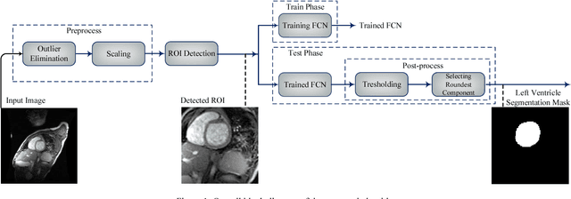
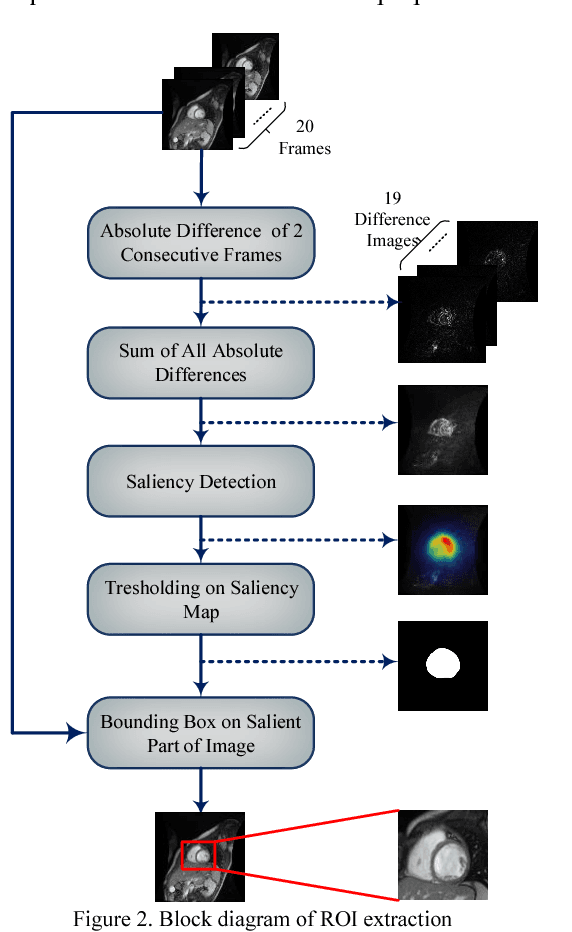

Abstract:Medical image analysis, especially segmenting a specific organ, has an important role in developing clinical decision support systems. In cardiac magnetic resonance (MR) imaging, segmenting the left and right ventricles helps physicians diagnose different heart abnormalities. There are challenges for this task, including the intensity and shape similarity between left ventricle and other organs, inaccurate boundaries and presence of noise in most of the images. In this paper we propose an automated method for segmenting the left ventricle in cardiac MR images. We first automatically extract the region of interest, and then employ it as an input of a fully convolutional network. We train the network accurately despite the small number of left ventricle pixels in comparison with the whole image. Thresholding on the output map of the fully convolutional network and selection of regions based on their roundness are performed in our proposed post-processing phase. The Dice score of our method reaches 87.24% by applying this algorithm on the York dataset of heart images.
Hand Gesture Recognition for Contactless Device Control in Operating Rooms
Nov 13, 2016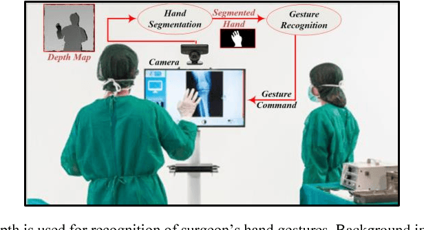
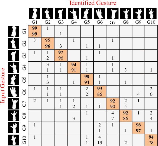

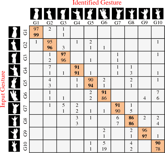
Abstract:Hand gesture is one of the most important means of touchless communication between human and machines. There is a great interest for commanding electronic equipment in surgery rooms by hand gesture for reducing the time of surgery and the potential for infection. There are challenges in implementation of a hand gesture recognition system. It has to fulfill requirements such as high accuracy and fast response. In this paper we introduce a system of hand gesture recognition based on a deep learning approach. Deep learning is known as an accurate detection model, but its high complexity prevents it from being fabricated as an embedded system. To cope with this problem, we applied some changes in the structure of our work to achieve low complexity. As a result, the proposed method could be implemented on a naive embedded system. Our experiments show that the proposed system results in higher accuracy while having less complexity in comparison with the existing comparable methods.
Extraction of Skin Lesions from Non-Dermoscopic Images Using Deep Learning
Sep 08, 2016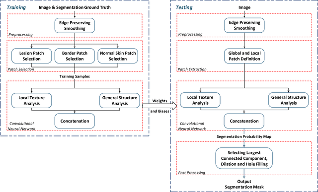
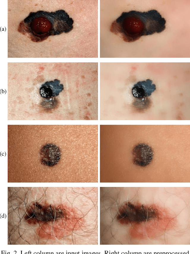
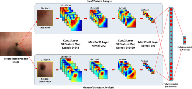
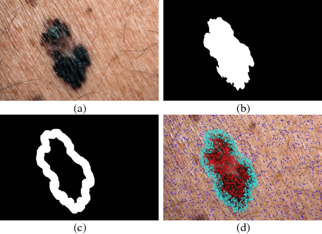
Abstract:Melanoma is amongst most aggressive types of cancer. However, it is highly curable if detected in its early stages. Prescreening of suspicious moles and lesions for malignancy is of great importance. Detection can be done by images captured by standard cameras, which are more preferable due to low cost and availability. One important step in computerized evaluation of skin lesions is accurate detection of lesion region, i.e. segmentation of an image into two regions as lesion and normal skin. Accurate segmentation can be challenging due to burdens such as illumination variation and low contrast between lesion and healthy skin. In this paper, a method based on deep neural networks is proposed for accurate extraction of a lesion region. The input image is preprocessed and then its patches are fed to a convolutional neural network (CNN). Local texture and global structure of the patches are processed in order to assign pixels to lesion or normal classes. A method for effective selection of training patches is used for more accurate detection of a lesion border. The output segmentation mask is refined by some post processing operations. The experimental results of qualitative and quantitative evaluations demonstrate that our method can outperform other state-of-the-art algorithms exist in the literature.
 Add to Chrome
 Add to Firefox
 Add to Edge