Mojtaba Akbari
Adaptive specular reflection detection and inpainting in colonoscopy video frames
Feb 23, 2018


Abstract:Colonoscopy video frames might be contaminated by bright spots with unsaturated values known as specular reflection. Detection and removal of such reflections could enhance the quality of colonoscopy images and facilitate diagnosis procedure. In this paper we propose a novel two-phase method for this purpose, consisting of detection and removal phases. In the detection phase, we employ both HSV and RGB color space information for segmentation of specular reflections. We first train a non-linear SVM for selecting a color space based on image statistical features extracted from each channel of the color spaces. Then, a cost function for detection of specular reflections is introduced. In the removal phase, we propose a two-step inpainting method which consists of appropriate replacement patch selection and removal of the blockiness effects. The proposed method is evaluated by testing on an available colonoscopy image database where accuracy and Dice score of 99.68% and 71.79% are achieved respectively.
Left Ventricle Segmentation in Cardiac MR Images Using Fully Convolutional Network
Feb 21, 2018


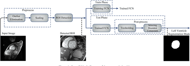
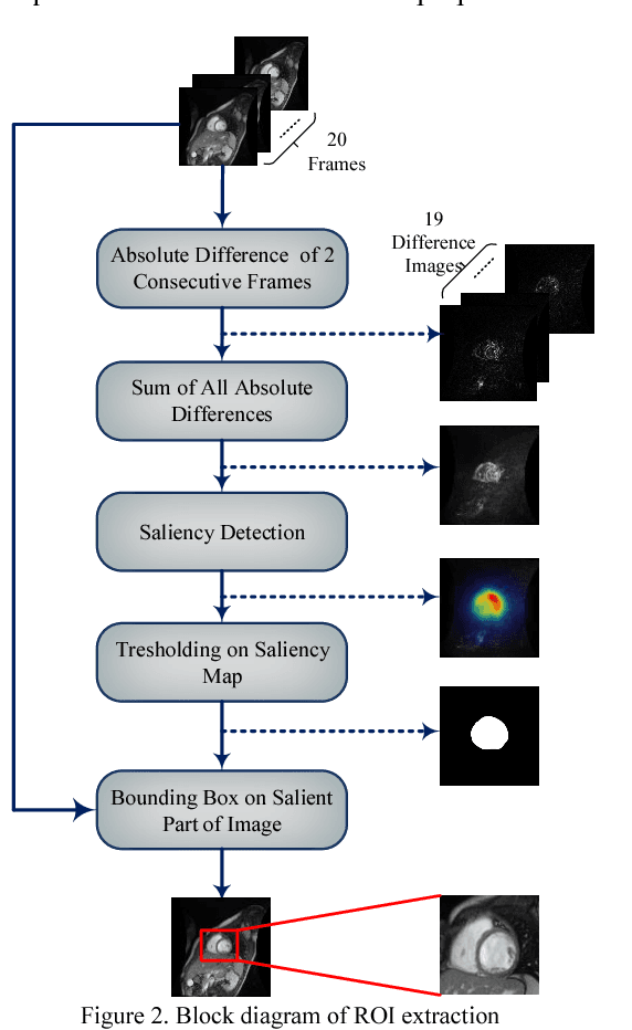
Abstract:Medical image analysis, especially segmenting a specific organ, has an important role in developing clinical decision support systems. In cardiac magnetic resonance (MR) imaging, segmenting the left and right ventricles helps physicians diagnose different heart abnormalities. There are challenges for this task, including the intensity and shape similarity between left ventricle and other organs, inaccurate boundaries and presence of noise in most of the images. In this paper we propose an automated method for segmenting the left ventricle in cardiac MR images. We first automatically extract the region of interest, and then employ it as an input of a fully convolutional network. We train the network accurately despite the small number of left ventricle pixels in comparison with the whole image. Thresholding on the output map of the fully convolutional network and selection of regions based on their roundness are performed in our proposed post-processing phase. The Dice score of our method reaches 87.24% by applying this algorithm on the York dataset of heart images.
 Add to Chrome
 Add to Firefox
 Add to Edge