Claudio Delrieux
Recursive Variational Autoencoders for 3D Blood Vessel Generative Modeling
Jun 17, 2025Abstract:Anatomical trees play an important role in clinical diagnosis and treatment planning. Yet, accurately representing these structures poses significant challenges owing to their intricate and varied topology and geometry. Most existing methods to synthesize vasculature are rule based, and despite providing some degree of control and variation in the structures produced, they fail to capture the diversity and complexity of actual anatomical data. We developed a Recursive variational Neural Network (RvNN) that fully exploits the hierarchical organization of the vessel and learns a low-dimensional manifold encoding branch connectivity along with geometry features describing the target surface. After training, the RvNN latent space can be sampled to generate new vessel geometries. By leveraging the power of generative neural networks, we generate 3D models of blood vessels that are both accurate and diverse, which is crucial for medical and surgical training, hemodynamic simulations, and many other purposes. These results closely resemble real data, achieving high similarity in vessel radii, length, and tortuosity across various datasets, including those with aneurysms. To the best of our knowledge, this work is the first to utilize this technique for synthesizing blood vessels.
VesselGPT: Autoregressive Modeling of Vascular Geometry
May 19, 2025Abstract:Anatomical trees are critical for clinical diagnosis and treatment planning, yet their complex and diverse geometry make accurate representation a significant challenge. Motivated by the latest advances in large language models, we introduce an autoregressive method for synthesizing anatomical trees. Our approach first embeds vessel structures into a learned discrete vocabulary using a VQ-VAE architecture, then models their generation autoregressively with a GPT-2 model. This method effectively captures intricate geometries and branching patterns, enabling realistic vascular tree synthesis. Comprehensive qualitative and quantitative evaluations reveal that our technique achieves high-fidelity tree reconstruction with compact discrete representations. Moreover, our B-spline representation of vessel cross-sections preserves critical morphological details that are often overlooked in previous' methods parameterizations. To the best of our knowledge, this work is the first to generate blood vessels in an autoregressive manner. Code, data, and trained models will be made available.
Deep learning and whole-brain networks for biomarker discovery: modeling the dynamics of brain fluctuations in resting-state and cognitive tasks
Dec 26, 2024Abstract:Background: Brain network models offer insights into brain dynamics, but the utility of model-derived bifurcation parameters as biomarkers remains underexplored. Objective: This study evaluates bifurcation parameters from a whole-brain network model as biomarkers for distinguishing brain states associated with resting-state and task-based cognitive conditions. Methods: Synthetic BOLD signals were generated using a supercritical Hopf brain network model to train deep learning models for bifurcation parameter prediction. Inference was performed on Human Connectome Project data, including both resting-state and task-based conditions. Statistical analyses assessed the separability of brain states based on bifurcation parameter distributions. Results: Bifurcation parameter distributions differed significantly across task and resting-state conditions ($p < 0.0001$ for all but one comparison). Task-based brain states exhibited higher bifurcation values compared to rest. Conclusion: Bifurcation parameters effectively differentiate cognitive and resting states, warranting further investigation as biomarkers for brain state characterization and neurological disorder assessment.
VesselVAE: Recursive Variational Autoencoders for 3D Blood Vessel Synthesis
Jul 07, 2023Abstract:We present a data-driven generative framework for synthesizing blood vessel 3D geometry. This is a challenging task due to the complexity of vascular systems, which are highly variating in shape, size, and structure. Existing model-based methods provide some degree of control and variation in the structures produced, but fail to capture the diversity of actual anatomical data. We developed VesselVAE, a recursive variational Neural Network that fully exploits the hierarchical organization of the vessel and learns a low-dimensional manifold encoding branch connectivity along with geometry features describing the target surface. After training, the VesselVAE latent space can be sampled to generate new vessel geometries. To the best of our knowledge, this work is the first to utilize this technique for synthesizing blood vessels. We achieve similarities of synthetic and real data for radius (.97), length (.95), and tortuosity (.96). By leveraging the power of deep neural networks, we generate 3D models of blood vessels that are both accurate and diverse, which is crucial for medical and surgical training, hemodynamic simulations, and many other purposes.
ErgoExplorer: Interactive Ergonomic Risk Assessment from Video Collections
Sep 07, 2022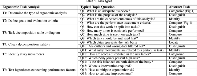
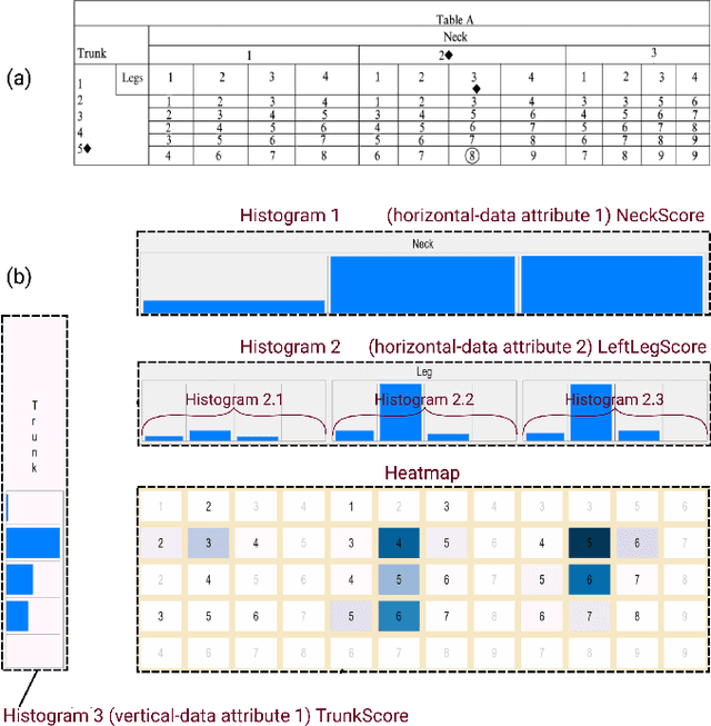
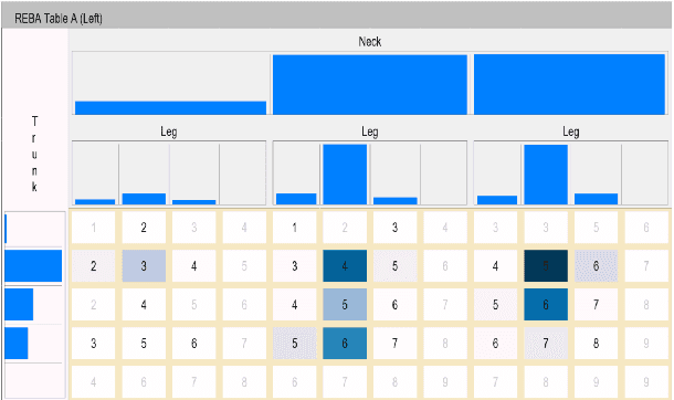
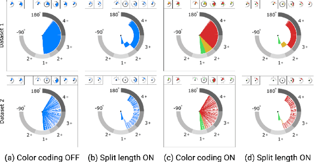
Abstract:Ergonomic risk assessment is now, due to an increased awareness, carried out more often than in the past. The conventional risk assessment evaluation, based on expert-assisted observation of the workplaces and manually filling in score tables, is still predominant. Data analysis is usually done with a focus on critical moments, although without the support of contextual information and changes over time. In this paper we introduce ErgoExplorer, a system for the interactive visual analysis of risk assessment data. In contrast to the current practice, we focus on data that span across multiple actions and multiple workers while keeping all contextual information. Data is automatically extracted from video streams. Based on carefully investigated analysis tasks, we introduce new views and their corresponding interactions. These views also incorporate domain-specific score tables to guarantee an easy adoption by domain experts. All views are integrated into ErgoExplorer, which relies on coordinated multiple views to facilitate analysis through interaction. ErgoExplorer makes it possible for the first time to examine complex relationships between risk assessments of individual body parts over long sessions that span multiple operations. The newly introduced approach supports analysis and exploration at several levels of detail, ranging from a general overview, down to inspecting individual frames in the video stream, if necessary. We illustrate the usefulness of the newly proposed approach applying it to several datasets.
Noise Reduction to Compute Tissue Mineral Density and Trabecular Bone Volume Fraction from Low Resolution QCT
Nov 04, 2020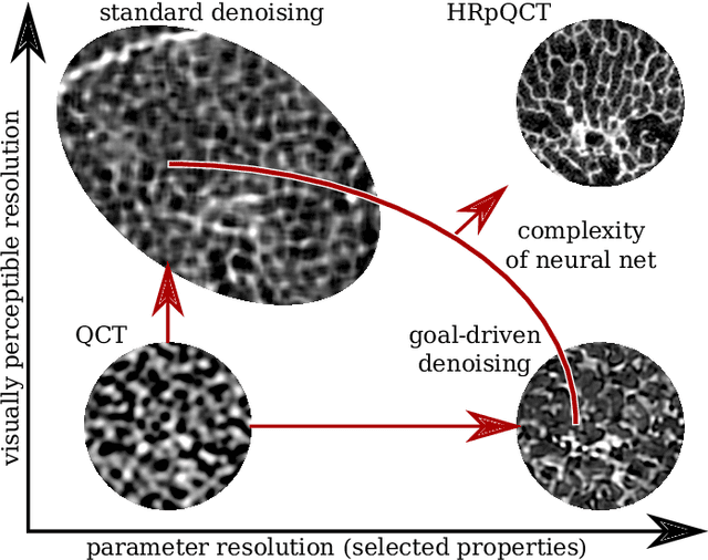
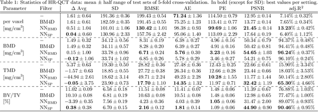


Abstract:We propose a 3D neural network with specific loss functions for quantitative computed tomography (QCT) noise reduction to compute micro-structural parameters such as tissue mineral density (TMD) and bone volume ratio (BV/TV) with significantly higher accuracy than using no or standard noise reduction filters. The vertebra-phantom study contained high resolution peripheral and clinical CT scans with simulated in vivo CT noise and nine repetitions of three different tube currents (100, 250 and 360 mAs). Five-fold cross validation was performed on 20466 purely spongy pairs of noisy and ground-truth patches. Comparison of training and test errors revealed high robustness against over-fitting. While not showing effects for the assessment of BMD and voxel-wise densities, the filter improved thoroughly the computation of TMD and BV/TV with respect to the unfiltered data. Root-mean-square and accuracy errors of low resolution TMD and BV/TV decreased to less than 17% of the initial values. Furthermore filtered low resolution scans revealed still more TMD- and BV/TV-relevant information than high resolution CT scans, either unfiltered or filtered with two state-of-the-art standard denoising methods. The proposed architecture is threshold and rotational invariant, applicable on a wide range of image resolutions at once, and likely serves for an accurate computation of further micro-structural parameters. Furthermore, it is less prone for over-fitting than neural networks that compute structural parameters directly. In conclusion, the method is potentially important for the diagnosis of osteoporosis and other bone diseases since it allows to assess relevant 3D micro-structural information from standard low exposure CT protocols such as 100 mAs and 120 kVp.
Generative Modelling of 3D in-silico Spongiosa with Controllable Micro-Structural Parameters
Sep 23, 2020
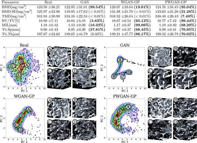
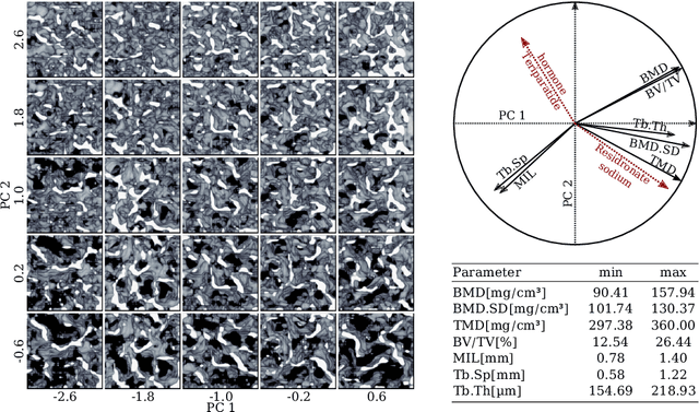
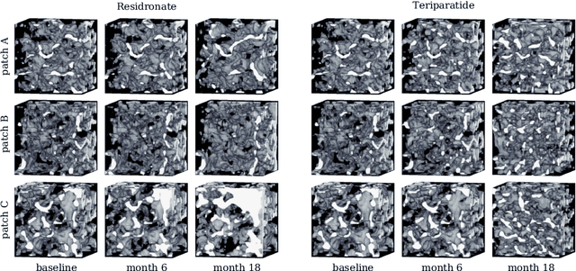
Abstract:Research in vertebral bone micro-structure generally requires costly procedures to obtain physical scans of real bone with a specific pathology under study, since no methods are available yet to generate realistic bone structures in-silico. Here we propose to apply recent advances in generative adversarial networks (GANs) to develop such a method. We adapted style-transfer techniques, which have been largely used in other contexts, in order to transfer style between image pairs while preserving its informational content. In a first step, we trained a volumetric generative model in a progressive manner using a Wasserstein objective and gradient penalty (PWGAN-GP) to create patches of realistic bone structure in-silico. The training set contained 7660 purely spongeous bone samples from twelve human vertebrae (T12 or L1) with isotropic resolution of 164um and scanned with a high resolution peripheral quantitative CT (Scanco XCT). After training, we generated new samples with tailored micro-structure properties by optimizing a vector z in the learned latent space. To solve this optimization problem, we formulated a differentiable goal function that leads to valid samples while compromising the appearance (content) with target 3D properties (style). Properties of the learned latent space effectively matched the data distribution. Furthermore, we were able to simulate the resulting bone structure after deterioration or treatment effects of osteoporosis therapies based only on expected changes of micro-structural parameters. Our method allows to generate a virtually infinite number of patches of realistic bone micro-structure, and thereby likely serves for the development of bone-biomarkers and to simulate bone therapies in advance.
SketchZooms: Deep multi-view descriptors for matching line drawings
Nov 29, 2019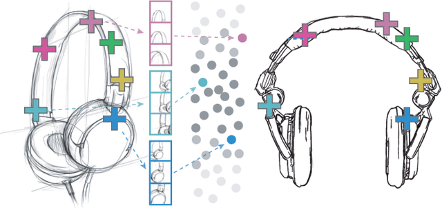


Abstract:Finding point-wise correspondences between images is a long-standing problem in computer vision. Corresponding sketch images is particularly challenging due to the varying nature of human style, projection distortions and viewport changes. In this paper we present a feature descriptor targeting line drawings learned from a 3D shape data set. Our descriptors are designed to locally match image pairs where the object of interest belongs to the same semantic category, yet still differ drastically in shape and projection angle. We build our descriptors by means of a Convolutional Neural Network (CNN) trained in a triplet fashion. The goal is to embed semantically similar anchor points close to one another, and to pull the embeddings of different points far apart. To learn the descriptors space, the network is fed with a succession of zoomed views from the input sketches. We have specifically crafted a data set of synthetic sketches using a non-photorealistic rendering algorithm over a large collection of part-based registered 3D models. Once trained, our network can generate descriptors for every pixel in an input image. Furthermore, our network is able to generalize well to unseen sketches hand-drawn by humans, outperforming state-of-the-art descriptors on the evaluated matching tasks. Our descriptors can be used to obtain sparse and dense correspondences between image pairs. We evaluate our method against a baseline of correspondences data collected from expert designers, in addition to comparisons with descriptors that have been proven effective in sketches. Finally, we demonstrate applications showing the usefulness of our multi-view descriptors.
 Add to Chrome
 Add to Firefox
 Add to Edge