Bart Bijnens
A Computational Pipeline for Advanced Analysis of 4D Flow MRI in the Left Atrium
May 14, 2025Abstract:The left atrium (LA) plays a pivotal role in modulating left ventricular filling, but our comprehension of its hemodynamics is significantly limited by the constraints of conventional ultrasound analysis. 4D flow magnetic resonance imaging (4D Flow MRI) holds promise for enhancing our understanding of atrial hemodynamics. However, the low velocities within the LA and the limited spatial resolution of 4D Flow MRI make analyzing this chamber challenging. Furthermore, the absence of dedicated computational frameworks, combined with diverse acquisition protocols and vendors, complicates gathering large cohorts for studying the prognostic value of hemodynamic parameters provided by 4D Flow MRI. In this study, we introduce the first open-source computational framework tailored for the analysis of 4D Flow MRI in the LA, enabling comprehensive qualitative and quantitative analysis of advanced hemodynamic parameters. Our framework proves robust to data from different centers of varying quality, producing high-accuracy automated segmentations (Dice $>$ 0.9 and Hausdorff 95 $<$ 3 mm), even with limited training data. Additionally, we conducted the first comprehensive assessment of energy, vorticity, and pressure parameters in the LA across a spectrum of disorders to investigate their potential as prognostic biomarkers.
Handling confounding variables in statistical shape analysis -- application to cardiac remodelling
Jul 28, 2020


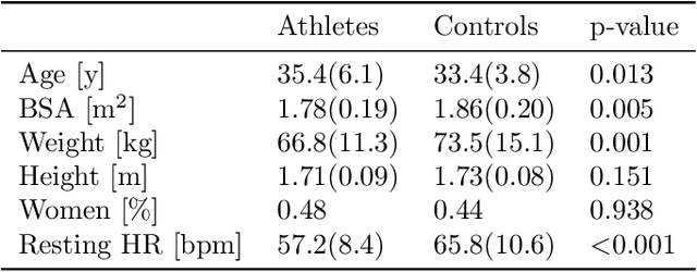
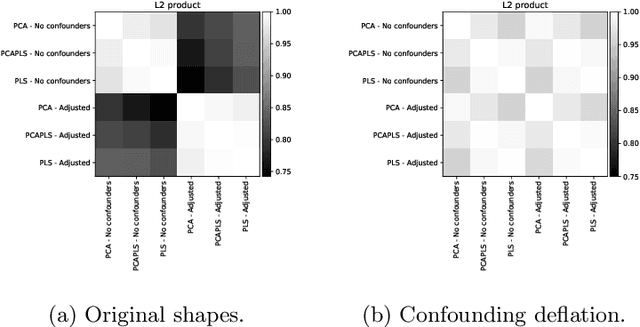
Abstract:Statistical shape analysis is a powerful tool to assess organ morphologies and find shape changes associated to a particular disease. However, imbalance in confounding factors, such as demographics might invalidate the analysis if not taken into consideration. Despite the methodological advances in the field, providing new methods that are able to capture complex and regional shape differences, the relationship between non-imaging information and shape variability has been overlooked. We present a linear statistical shape analysis framework that finds shape differences unassociated to a controlled set of confounding variables. It includes two confounding correction methods: confounding deflation and adjustment. We applied our framework to a cardiac magnetic resonance imaging dataset, consisting of the cardiac ventricles of 89 triathletes and 77 controls, to identify cardiac remodelling due to the practice of endurance exercise. To test robustness to confounders, subsets of this dataset were generated by randomly removing controls with low body mass index, thus introducing imbalance. The analysis of the whole dataset indicates an increase of ventricular volumes and myocardial mass in athletes, which is consistent with the clinical literature. However, when confounders are not taken into consideration no increase of myocardial mass is found. Using the downsampled datasets, we find that confounder adjustment methods are needed to find the real remodelling patterns in imbalanced datasets.
Volumetric parcellation of the right ventricle for regional geometric and functional assessment
Mar 18, 2020


Abstract:In clinical practice, assessment of right ventricle (RV) is primarily done through its global volume, given it is a standardised measurement, and has a good reproducibility in 3D modalities such as MRI and 3D echocardiography. However, many illness produce regionalchanges and therefore a local analysis could provide a better tool for understanding and diagnosis of illnesses. Current regional clinical measurements are 2D linear dimensions, and suffer of low reproducibility due to the difficulty to identify landmarks in the RV, specially in echocardiographic images due to its noise and artefacts. We proposed an automatic method for parcellating the RV cavity and compute regional volumes and ejection fractions in three regions: apex, inlet and outflow. We tested the reproducibility in 3D echocardiographic images. We also present a synthetic mesh-deformation method to generate a groundtruth dataset for validating the ability of the method to capture different types of remodelling. Results showed an acceptable intra-observer reproduciblity (<10%) but a higher inter-observer(>10%). The synthetic dataset allowed to identify that the method was capable of assessing global dilatations, and local dilatations in the circumferential direction but not longitudinal elongations
 Add to Chrome
 Add to Firefox
 Add to Edge