Balázs Gulyás
Contrastive Graph Pooling for Explainable Classification of Brain Networks
Jul 07, 2023


Abstract:Functional magnetic resonance imaging (fMRI) is a commonly used technique to measure neural activation. Its application has been particularly important in identifying underlying neurodegenerative conditions such as Parkinson's, Alzheimer's, and Autism. Recent analysis of fMRI data models the brain as a graph and extracts features by graph neural networks (GNNs). However, the unique characteristics of fMRI data require a special design of GNN. Tailoring GNN to generate effective and domain-explainable features remains challenging. In this paper, we propose a contrastive dual-attention block and a differentiable graph pooling method called ContrastPool to better utilize GNN for brain networks, meeting fMRI-specific requirements. We apply our method to 5 resting-state fMRI brain network datasets of 3 diseases and demonstrate its superiority over state-of-the-art baselines. Our case study confirms that the patterns extracted by our method match the domain knowledge in neuroscience literature, and disclose direct and interesting insights. Our contributions underscore the potential of ContrastPool for advancing the understanding of brain networks and neurodegenerative conditions.
3D Deep Learning on Medical Images: A Review
Apr 01, 2020


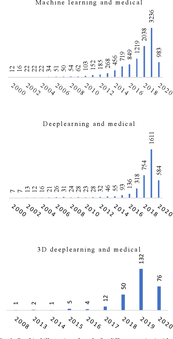
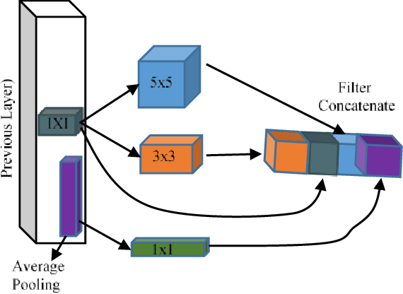
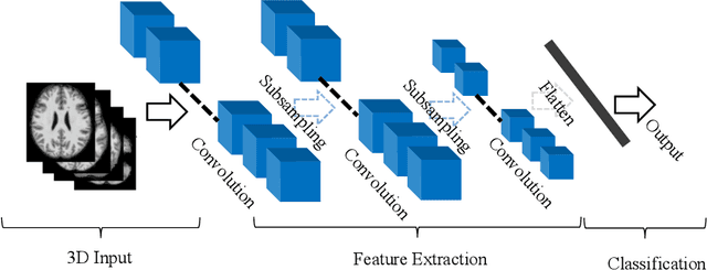
Abstract:The rapid advancements in machine learning, graphics processing technologies and availability of medical imaging data has led to a rapid increase in use of machine learning models in the medical domain. This was exacerbated by the rapid advancements in convolutional neural network (CNN) based architectures, which were adopted by the medical imaging community to assist clinicians in disease diagnosis. Since the grand success of AlexNet in 2012, CNNs have been increasingly used in medical image analysis to improve the efficiency of human clinicians. In recent years, three-dimensional (3D) CNNs have been employed for analysis of medical images. In this paper, we trace the history of how the 3D CNN was developed from its machine learning roots, brief mathematical description of 3D CNN and the preprocessing steps required for medical images before feeding them to 3D CNNs. We review the significant research in the field of 3D medical imaging analysis using 3D CNNs (and its variants) in different medical areas such as classification, segmentation, detection, and localization. We conclude by discussing the challenges associated with the use of 3D CNNs in the medical imaging domain (and the use of deep learning models, in general) and possible future trends in the field.
 Add to Chrome
 Add to Firefox
 Add to Edge