Ascanio Tridente
CXR-Net: An Encoder-Decoder-Encoder Multitask Deep Neural Network for Explainable and Accurate Diagnosis of COVID-19 pneumonia with Chest X-ray Images
Oct 20, 2021


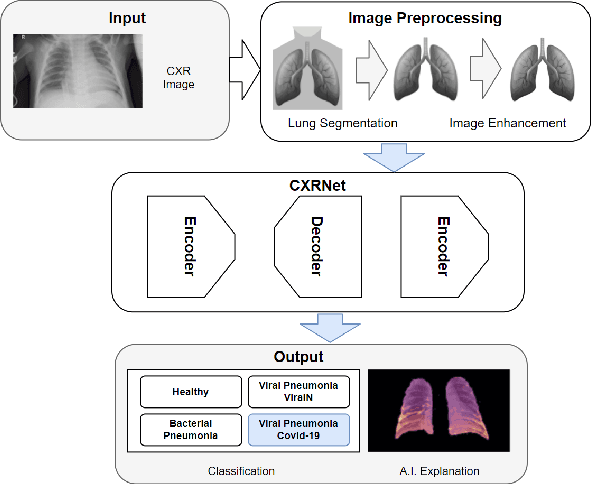
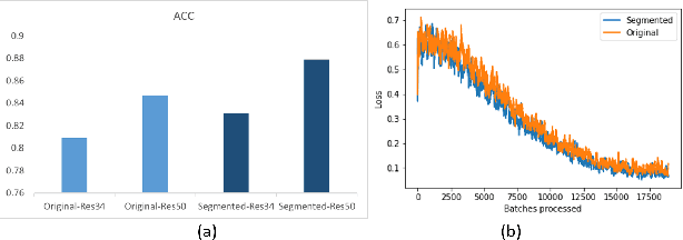
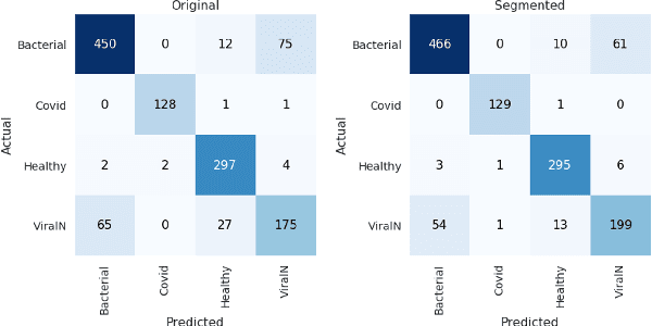
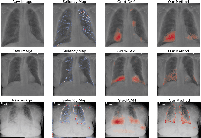
Abstract:Accurate and rapid detection of COVID-19 pneumonia is crucial for optimal patient treatment. Chest X-Ray (CXR) is the first line imaging test for COVID-19 pneumonia diagnosis as it is fast, cheap and easily accessible. Inspired by the success of deep learning (DL) in computer vision, many DL-models have been proposed to detect COVID-19 pneumonia using CXR images. Unfortunately, these deep classifiers lack the transparency in interpreting findings, which may limit their applications in clinical practice. The existing commonly used visual explanation methods are either too noisy or imprecise, with low resolution, and hence are unsuitable for diagnostic purposes. In this work, we propose a novel explainable deep learning framework (CXRNet) for accurate COVID-19 pneumonia detection with an enhanced pixel-level visual explanation from CXR images. The proposed framework is based on a new Encoder-Decoder-Encoder multitask architecture, allowing for both disease classification and visual explanation. The method has been evaluated on real world CXR datasets from both public and private data sources, including: healthy, bacterial pneumonia, viral pneumonia and COVID-19 pneumonia cases The experimental results demonstrate that the proposed method can achieve a satisfactory level of accuracy and provide fine-resolution classification activation maps for visual explanation in lung disease detection. The Average Accuracy, the Precision, Recall and F1-score of COVID-19 pneumonia reached 0.879, 0.985, 0.992 and 0.989, respectively. We have also found that using lung segmented (CXR) images can help improve the performance of the model. The proposed method can provide more detailed high resolution visual explanation for the classification decision, compared to current state-of-the-art visual explanation methods and has a great potential to be used in clinical practice for COVID-19 pneumonia diagnosis.
 Add to Chrome
 Add to Firefox
 Add to Edge