Computer-Aided Extraction of Select MRI Markers of Cerebral Small Vessel Disease: A Systematic Review
Paper and Code
Apr 04, 2022


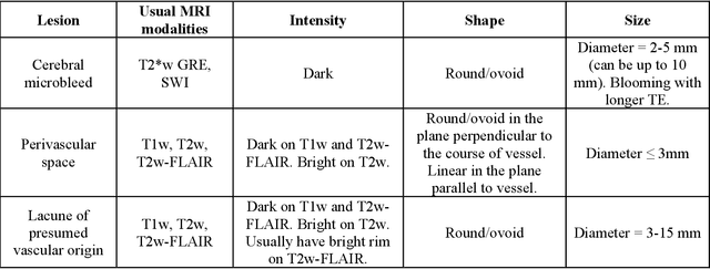
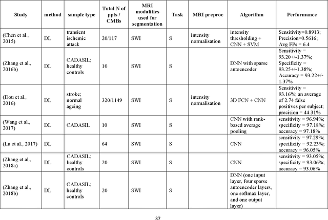
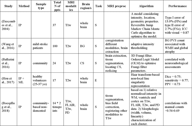
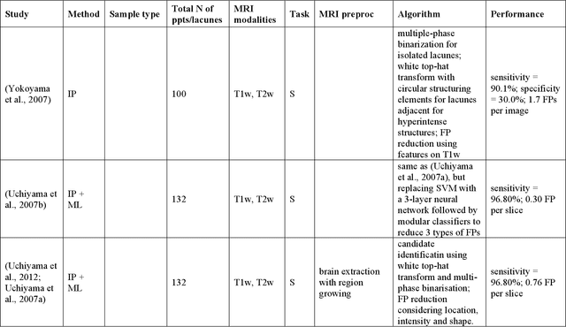
Cerebral small vessel disease (CSVD) is a major vascular contributor to cognitive impairment in ageing, including dementias. Imaging remains the most promising method for in vivo studies of CSVD. To replace the subjective and laborious visual rating approaches, emerging studies have applied state-of-the-art artificial intelligence to extract imaging biomarkers of CSVD from MRI scans. We aimed to summarise published computer-aided methods to examine three imaging biomarkers of CSVD, namely cerebral microbleeds (CMB), dilated perivascular spaces (PVS), and lacunes of presumed vascular origin. Seventy-one classical image processing, classical machine learning, and deep learning studies were identified. CMB and PVS have been better studied, compared to lacunes. While good performance metrics have been achieved in local test datasets, there have not been generalisable pipelines validated in different research or clinical cohorts. Transfer learning and weak supervision techniques have been applied to accommodate the limitations in training data. Future studies could consider pooling data from multiple sources to increase diversity, and validating the performance of the methods using both image processing metrics and associations with clinical measures.
 Add to Chrome
 Add to Firefox
 Add to Edge