Yin Yin
Cerebrovascular Segmentation via Vessel Oriented Filtering Network
Oct 17, 2022


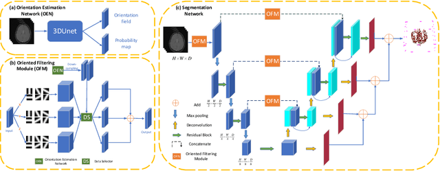
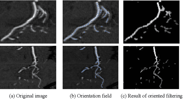
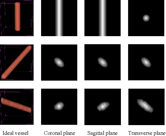
Abstract:Accurate cerebrovascular segmentation from Magnetic Resonance Angiography (MRA) and Computed Tomography Angiography (CTA) is of great significance in diagnosis and treatment of cerebrovascular pathology. Due to the complexity and topology variability of blood vessels, complete and accurate segmentation of vascular network is still a challenge. In this paper, we proposed a Vessel Oriented Filtering Network (VOF-Net) which embeds domain knowledge into the convolutional neural network. We design oriented filters for blood vessels according to vessel orientation field, which is obtained by orientation estimation network. Features extracted by oriented filtering are injected into segmentation network, so as to make use of the prior information that the blood vessels are slender and curved tubular structure. Experimental results on datasets of CTA and MRA show that the proposed method is effective for vessel segmentation, and embedding the specific vascular filter improves the segmentation performance.
Transferring Learned Microcalcification Group Detection from 2D Mammography to 3D Digital Breast Tomosynthesis Using a Hierarchical Model and Scope-based Normalization Features
Mar 18, 2016


Abstract:A novel hierarchical model is introduced to solve a general problem of detecting groups of similar objects. Under this model, detection of groups is performed in hierarchically organized layers while each layer represents a scope for target objects. The processing of these layers involves sequential extraction of appearance features for an individual object, consistency measurement features for nearby objects, and finally the distribution features for all objects within the group. Using the concept of scope-based normalization, the extracted features not only enhance local contrast of an individual object, but also provide consistent characterization for all related objects. As an example, a microcalcification group detection system for 2D mammography was developed, and then the learned model was transferred to 3D digital breast tomosynthesis without any retraining or fine-tuning. The detection system demonstrated state-of-the-art performance and detected 96% of cancerous lesions at the rate of 1.2 false positives per volume as measured on an independent tomosynthesis test set.
 Add to Chrome
 Add to Firefox
 Add to Edge