Xuan Lei
Image Registration with Averaging Network and Edge-Based Loss for Low-SNR Cardiac MRI
Sep 04, 2024Abstract:Purpose: To perform image registration and averaging of multiple free-breathing single-shot cardiac images, where the individual images may have a low signal-to-noise ratio (SNR). Methods: To address low SNR encountered in single-shot imaging, especially at low field strengths, we propose a fast deep learning (DL)-based image registration method, called Averaging Morph with Edge Detection (AiM-ED). AiM-ED jointly registers multiple noisy source images to a noisy target image and utilizes a noise-robust pre-trained edge detector to define the training loss. We validate AiM-ED using synthetic late gadolinium enhanced (LGE) imaging data from the MR extended cardiac-torso (MRXCAT) phantom and retrospectively undersampled single-shot data from healthy subjects (24 slices) and patients (5 slices) under various levels of added noise. Additionally, we demonstrate the clinical feasibility of AiM-ED by applying it to prospectively undersampled data from patients (6 slices) scanned at a 0.55T scanner. Results: Compared to a traditional energy-minimization-based image registration method and DL-based VoxelMorph, images registered using AiM-ED exhibit higher values of recovery SNR and three perceptual image quality metrics. An ablation study shows the benefit of both jointly processing multiple source images and using an edge map in AiM-ED. Conclusion: For single-shot LGE imaging, AiM-ED outperforms existing image registration methods in terms of image quality. With fast inference, minimal training data requirements, and robust performance at various noise levels, AiM-ED has the potential to benefit single-shot CMR applications.
Surface Coil Intensity Correction for MRI
Dec 01, 2023


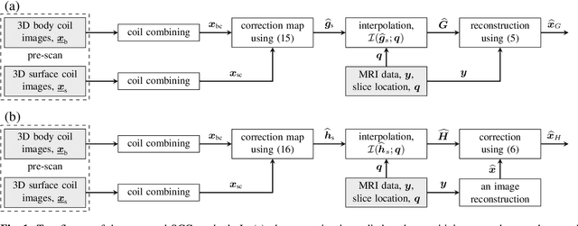
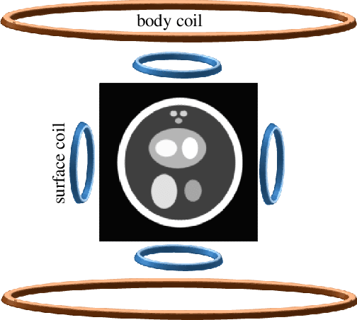
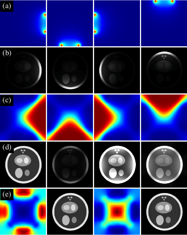
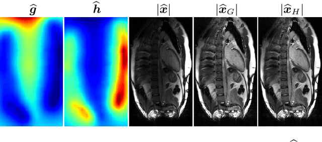
Abstract:Modern MRI scanners utilize one or more arrays of small receive-only coils to collect k-space data. The sensitivity maps of the coils, when estimated using traditional methods, differ from the true sensitivity maps, which are generally unknown. Consequently, the reconstructed MR images exhibit undesired spatial variation in intensity. These intensity variations can be at least partially corrected using pre-scan data. In this work, we propose an intensity correction method that utilizes pre-scan data. For demonstration, we apply our method to a digital phantom, as well as to cardiac MRI data collected from a commercial scanner by Siemens Healthineers. The code is available at https://github.com/OSU-MR/SCC.
 Add to Chrome
 Add to Firefox
 Add to Edge