Vismay Agrawal
Structurally aware bidirectional unpaired image to image translation between CT and MR
Jun 05, 2020
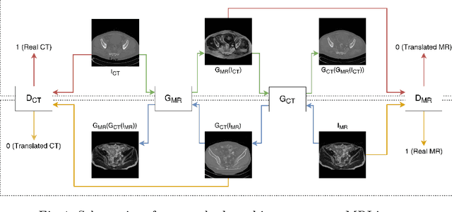
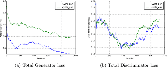

Abstract:Magnetic Resonance (MR) Imaging and Computed Tomography (CT) are the primary diagnostic imaging modalities quite frequently used for surgical planning and analysis. A general problem with medical imaging is that the acquisition process is quite expensive and time-consuming. Deep learning techniques like generative adversarial networks (GANs) can help us to leverage the possibility of an image to image translation between multiple imaging modalities, which in turn helps in saving time and cost. These techniques will help to conduct surgical planning under CT with the feedback of MRI information. While previous studies have shown paired and unpaired image synthesis from MR to CT, image synthesis from CT to MR still remains a challenge, since it involves the addition of extra tissue information. In this manuscript, we have implemented two different variations of Generative Adversarial Networks exploiting the cycling consistency and structural similarity between both CT and MR image modalities on a pelvis dataset, thus facilitating a bidirectional exchange of content and style between these image modalities. The proposed GANs translate the input medical images by different mechanisms, and hence generated images not only appears realistic but also performs well across various comparison metrics, and these images have also been cross verified with a radiologist. The radiologist verification has shown that slight variations in generated MR and CT images may not be exactly the same as their true counterpart but it can be used for medical purposes.
Enhanced Optic Disk and Cup Segmentation with Glaucoma Screening from Fundus Images using Position encoded CNNs
Sep 14, 2018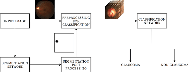
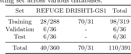
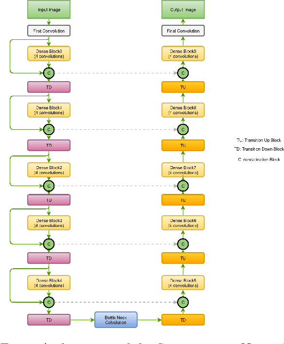

Abstract:In this manuscript, we present a robust method for glaucoma screening from fundus images using an ensemble of convolutional neural networks (CNNs). The pipeline comprises of first segmenting the optic disk and optic cup from the fundus image, then extracting a patch centered around the optic disk and subsequently feeding to the classification network to differentiate the image as diseased or healthy. In the segmentation network, apart from the image, we make use of spatial co-ordinate (X \& Y) space so as to learn the structure of interest better. The classification network is composed of a DenseNet201 and a ResNet18 which were pre-trained on a large cohort of natural images. On the REFUGE validation data (n=400), the segmentation network achieved a dice score of 0.88 and 0.64 for optic disc and optic cup respectively. For the tasking differentiating images affected with glaucoma from healthy images, the area under the ROC curve was observed to be 0.85.
 Add to Chrome
 Add to Firefox
 Add to Edge