Tom R. L. Kimpe
On anthropomorphic decision making in a model observer
Jun 30, 2015Abstract:By analyzing human readers' performance in detecting small round lesions in simulated digital breast tomosynthesis background in a location known exactly scenario, we have developed a model observer that is a better predictor of human performance with different levels of background complexity (i.e., anatomical and quantum noise). Our analysis indicates that human observers perform a lesion detection task by combining a number of sub-decisions, each an indicator of the presence of a lesion in the image stack. This is in contrast to a channelized Hotelling observer, where the detection task is conducted holistically by thresholding a single decision variable, made from an optimally weighted linear combination of channels. However, it seems that the sub-par performance of human readers compared to the CHO cannot be fully explained by their reliance on sub-decisions, or perhaps we do not consider a sufficient number of sub-decisions. To bridge the gap between the performances of human readers and the model observer based upon sub-decisions, we use an additive noise model, the power of which is modulated with the level of background complexity. The proposed model observer better predicts the fast drop in human detection performance with background complexity.
Aging display's effect on interpretation of digital pathology slides
Jun 30, 2015Abstract:It is our conjecture that the variability of colors in a pathology image effects the interpretation of pathology cases, whether it is diagnostic accuracy, diagnostic confidence, or workflow efficiency. In this paper, digital pathology images are analyzed to quantify the perceived difference in color that occurs due to display aging, in particular a change in the maximum luminance, white point, and color gamut. The digital pathology images studied include diagnostically important features, such as the conspicuity of nuclei. Three different display aging models are applied to images: aging of luminance & chrominance, aging of chrominance only, and a stabilized luminance & chrominance (i.e., no aging). These display models and images are then used to compare conspicuity of nuclei using CIE deltaE2000, a perceptual color difference metric. The effect of display aging using these display models and images is further analyzed through a human reader study designed to quantify the effects from a clinical perspective. Results from our reader study indicate significant impact of aged displays on workflow as well as diagnosis as follow. As compared to the originals (no-aging), slides with the effect of aging simulated were significantly more difficult to read (p-value of 0.0005) and took longer to score (p-value of 0.02). Moreover, luminance+chrominance aging significantly reduced inter-session percent agreement of diagnostic scores (p-value of 0.0418).
It is hard to see a needle in a haystack: Modeling contrast masking effect in a numerical observer
Aug 05, 2014


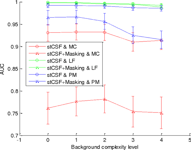
Abstract:Within the framework of a virtual clinical trial for breast imaging, we aim to develop numerical observers that follow the same detection performance trends as those of a typical human observer. In our prior work, we showed that by including spatiotemporal contrast sensitivity function (stCSF) of human visual system (HVS) in a multi-slice channelized Hotelling observer (msCHO), we can correctly predict trends of a typical human observer performance with the viewing parameters of browsing speed, viewing distance and contrast. In this work we further improve our numerical observer by modeling contrast masking. After stCSF, contrast masking is the second most prominent property of HVS and it refers to the fact that the presence of one signal affects the visibility threshold for another signal. Our results indicate that the improved numerical observer better predicts changes in detection performance with background complexity.
Development and evaluation of a 3D model observer with nonlinear spatiotemporal contrast sensitivity
Mar 24, 2014Abstract:We investigate improvements to our 3D model observer with the goal of better matching human observer performance as a function of viewing distance, effective contrast, maximum luminance, and browsing speed. Two nonlinear methods of applying the human contrast sensitivity function (CSF) to a 3D model observer are proposed, namely the Probability Map (PM) and Monte Carlo (MC) methods. In the PM method, the visibility probability for each frequency component of the image stack, p, is calculated taking into account Barten's spatiotemporal CSF, the component modulation, and the human psychometric function. The probability p is considered to be equal to the perceived amplitude of the frequency component and thus can be used by a traditional model observer (e.g., LG-msCHO) in the space-time domain. In the MC method, each component is randomly kept with probability p or discarded with 1-p. The amplitude of the retained components is normalized to unity. The methods were tested using DBT stacks of an anthropomorphic breast phantom processed in a comprehensive simulation pipeline. Our experiments indicate that both the PM and MC methods yield results that match human observer performance better than the linear filtering method as a function of viewing distance, effective contrast, maximum luminance, and browsing speed.
Integration of spatio-temporal contrast sensitivity with a multi-slice channelized Hotelling observer
Apr 04, 2013Abstract:Barten's model of spatio-temporal contrast sensitivity function of human visual system is embedded in a multi-slice channelized Hotelling observer. This is done by 3D filtering of the stack of images with the spatio-temporal contrast sensitivity function and feeding the result (i.e., the perceived image stack) to the multi-slice channelized Hotelling observer. The proposed procedure of considering spatio-temporal contrast sensitivity function is generic in the sense that it can be used with observers other than multi-slice channelized Hotelling observer. Detection performance of the new observer in digital breast tomosynthesis is measured in a variety of browsing speeds, at two spatial sampling rates, using computer simulations. Our results show a peak in detection performance in mid browsing speeds. We compare our results to those of a human observer study reported earlier (I. Diaz et al. SPIE MI 2011). The effects of display luminance, contrast and spatial sampling rate, with and without considering foveal vision, are also studied. Reported simulations are conducted with real digital breast tomosynthesis image stacks, as well as stacks from an anthropomorphic software breast phantom (P. Bakic et al. Med Phys. 2011). Lesion cases are simulated by inserting single micro-calcifications or masses. Limitations of our methods and ways to improve them are discussed.
 Add to Chrome
 Add to Firefox
 Add to Edge