Tim Laibacher
On the Evaluation and Real-World Usage Scenarios of Deep Vessel Segmentation for Funduscopy
Sep 12, 2019


Abstract:We identify and address three research gaps in the field of vessel segmentation for funduscopy. The first focuses on the task of inference on high-resolution fundus images for which only a limited set of ground-truth data is publicly available. Notably, we highlight that simple rescaling and padding or cropping of lower resolution datasets is surprisingly effective. Additionally we explore the effectiveness of semi-supervised learning for better domain adaptation. Our results show competitive performance on a set of common public retinal vessel datasets using a small and light-weight neural network. For HRF, the only very high-resolution dataset currently available, we reach new state-of-the-art performance by solely relying on training images from lower-resolution datasets. The second topic concerns evaluation metrics. We investigate the variability of the F1-score on the existing datasets and report results for recent SOTA architectures. Our evaluation show that most SOTA results are actually comparable to each other in performance. Last, we address the issue of reproducibility by open-sourcing our complete pipeline.
M2U-Net: Effective and Efficient Retinal Vessel Segmentation for Resource-Constrained Environments
Dec 03, 2018


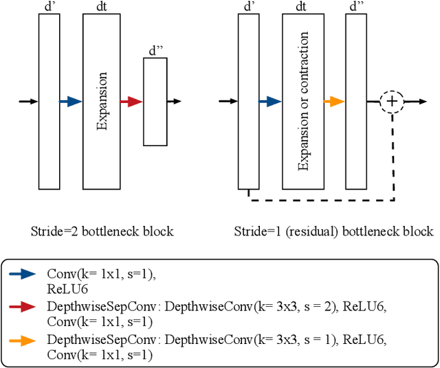
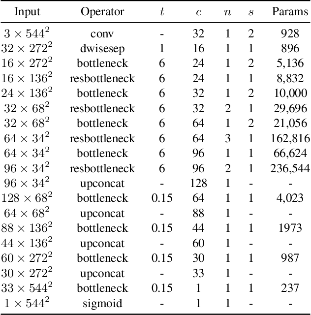
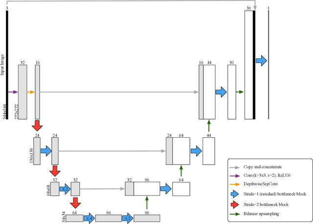
Abstract:In this paper, we present a novel neural network architecture for retinal vessel segmentation that improves over the state of the art on two benchmark datasets, is the first to run in real time on high resolution images, and its small memory and processing requirements make it deployable in mobile and embedded systems. The M2U-Net has a new encoder-decoder architecture that is inspired by the U-Net. It adds pretrained components of MobileNetV2 in the encoder part and novel contractive bottleneck blocks in the decoder part that, combined with bilinear upsampling, drastically reduce the parameter count to 0.55M compared to 31.03M in the original U-Net. We have evaluated its performance against a wide body of previously published results on three public datasets. On two of them, the M2U-Net achieves new state-of-the-art performance by a considerable margin. When implemented on a GPU, our method is the first to achieve real-time inference speeds on high-resolution fundus images. We also implemented our proposed network on an ARM-based embedded system where it segments images in between 0.6 and 15 sec, depending on the resolution. Thus, the M2U-Net enables a number of applications of retinal vessel structure extraction, such as early diagnosis of eye diseases, retinal biometric authentication systems, and robot assisted microsurgery.
 Add to Chrome
 Add to Firefox
 Add to Edge