Shouvik Biswas
Embedding of Blink Frequency in Electrooculography Signal using Difference Expansion based Reversible Watermarking Technique
Mar 09, 2013


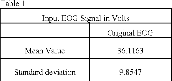
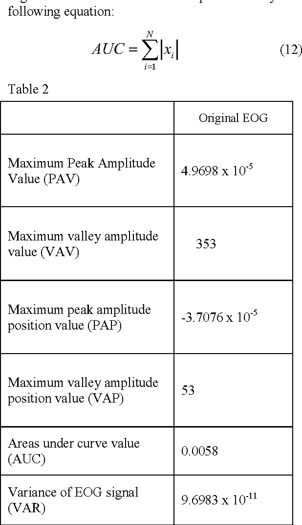
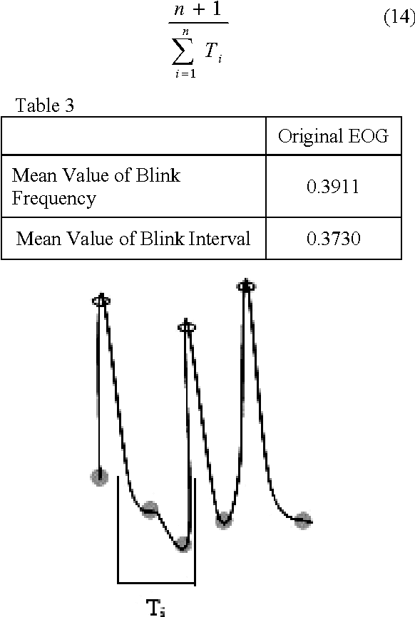
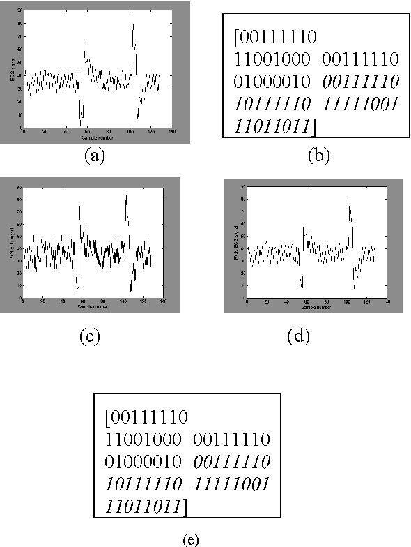
Abstract:In the past few years, like other fields, rapid expansion of digitization and globalization has influenced the medical field as well. For progress of diagnostic results most of the reputed hospitals and diagnostic centres all over the world have started exchanging medical information. In this proposed method, the calculated diagnostic parametric values of the original Electrooculography (EOG) signal are embedded as a watermark by using Difference Expansion (DE) algorithm based reversible watermarking technique. The extracted watermark provides the required parametric values at the recipient end without any post computation of the recovered EOG signal. By computing the parametric values from the recovered signal, the integrity of the extracted watermark can be validated. The time domain features of EOG signal are calculated for the generation of watermark. In the current work, various features are studied and two major features related to blink frequency are used to generate the watermark. The high Signal to Noise Ratio (SNR) and the Bit Error Rate (BER) claim the robustness of the proposed method.
* 6 Pages, 3 Figures, 4 Tables
Wavelet Based QRS Complex Detection of ECG Signal
Sep 07, 2012


Abstract:The Electrocardiogram (ECG) is a sensitive diagnostic tool that is used to detect various cardiovascular diseases by measuring and recording the electrical activity of the heart in exquisite detail. A wide range of heart condition is determined by thorough examination of the features of the ECG report. Automatic extraction of time plane features is important for identification of vital cardiac diseases. This paper presents a multi-resolution wavelet transform based system for detection 'P', 'Q', 'R', 'S', 'T' peaks complex from original ECG signal. 'R-R' time lapse is an important minutia of the ECG signal that corresponds to the heartbeat of the concerned person. Abrupt increase in height of the 'R' wave or changes in the measurement of the 'R-R' denote various anomalies of human heart. Similarly 'P-P', 'Q-Q', 'S-S', 'T-T' also corresponds to different anomalies of heart and their peak amplitude also envisages other cardiac diseases. In this proposed method the 'PQRST' peaks are marked and stored over the entire signal and the time interval between two consecutive 'R' peaks and other peaks interval are measured to detect anomalies in behavior of heart, if any. The peaks are achieved by the composition of Daubeheissub bands wavelet of original ECG signal. The accuracy of the 'PQRST' complex detection and interval measurement is achieved up to 100% with high exactitude by processing and thresholding the original ECG signal.
* 5 pages, 8 figures, ISSN: 2248-9622
 Add to Chrome
 Add to Firefox
 Add to Edge