Nicoló Savioli
A Hybrid Approach Between Adversarial Generative Networks and Actor-Critic Policy Gradient for Low Rate High-Resolution Image Compression
Jun 13, 2019


Abstract:Image compression is an essential approach for decreasing the size in bytes of the image without deteriorating the quality of it. Typically, classic algorithms are used but recently deep-learning has been successfully applied. In this work, is presented a deep super-resolution work-flow for image compression that maps low-resolution JPEG image to the high-resolution. The pipeline consists of two components: first, an encoder-decoder neural network learns how to transform the downsampling JPEG images to high resolution. Second, a combination between Generative Adversarial Networks (GANs) and reinforcement learning Actor-Critic (A3C) loss pushes the encoder-decoder to indirectly maximize High Peak Signal-to-Noise Ratio (PSNR). Although PSNR is a fully differentiable metric, this work opens the doors to new solutions for maximizing non-differential metrics through an end-to-end approach between encoder-decoder networks and reinforcement learning policy gradient methods.
A Generative Adversarial Model for Right Ventricle Segmentation
Sep 27, 2018
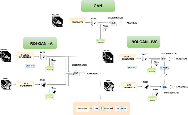
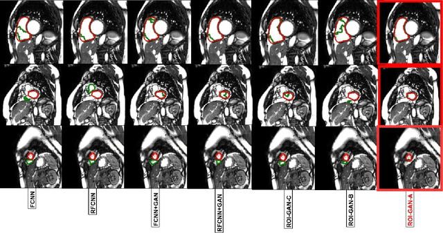

Abstract:The clinical management of several cardiovascular conditions, such as pulmonary hypertension, require the assessment of the right ventricular (RV) function. This work addresses the fully automatic and robust access to one of the key RV biomarkers, its ejection fraction, from the gold standard imaging modality, MRI. The problem becomes the accurate segmentation of the RV blood pool from cine MRI sequences. This work proposes a solution based on Fully Convolutional Neural Networks (FCNN), where our first contribution is the optimal combination of three concepts (the convolution Gated Recurrent Units (GRU), the Generative Adversarial Networks (GAN), and the L1 loss function) that achieves an improvement of 0.05 and 3.49 mm in Dice Index and Hausdorff Distance respectively with respect to the baseline FCNN. This improvement is then doubled by our second contribution, the ROI-GAN, that sets two GANs to cooperate working at two fields of view of the image, its full resolution and the region of interest (ROI). Our rationale here is to better guide the FCNN learning by combining global (full resolution) and local Region Of Interest (ROI) features. The study is conducted in a large in-house dataset of $\sim$ 23.000 segmented MRI slices, and its generality is verified in a publicly available dataset.
V-FCNN: Volumetric Fully Convolution Neural Network For Automatic Atrial Segmentation
Sep 27, 2018


Abstract:Atrial Fibrillation (AF) is a common electro-physiological cardiac disorder that causes changes in the anatomy of the atria. A better characterization of these changes is desirable for the definition of clinical biomarkers, furthermore, thus there is a need for its fully automatic segmentation from clinical images. In this work, we present an architecture based on 3D-convolution kernels, a Volumetric Fully Convolution Neural Network (V-FCNN), able to segment the entire volume in a one-shot, and consequently integrate the implicit spatial redundancy present in high-resolution images. A loss function based on the mixture of both Mean Square Error (MSE) and Dice Loss (DL) is used, in an attempt to combine the ability to capture the bulk shape as well as the reduction of local errors products by over-segmentation. Results demonstrate a reasonable performance in the middle region of the atria along with the impact of the challenges of capturing the variability of the pulmonary veins or the identification of the valve plane that separates the atria to the ventricle. A final dice of $92.5\%$ in $54$ patients ($4752$ atria test slices in total) is shown.
Automated segmentation on the entire cardiac cycle using a deep learning work-flow
Aug 31, 2018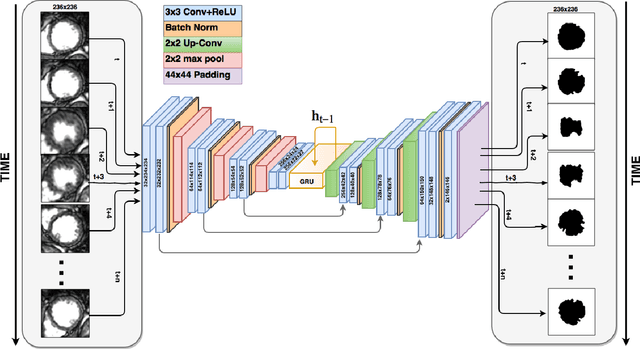
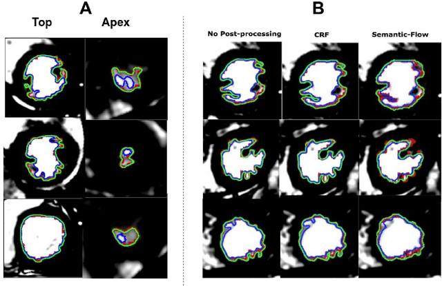
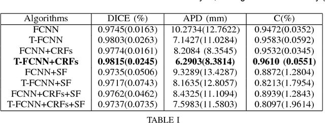
Abstract:The segmentation of the left ventricle (LV) from CINE MRI images is essential to infer important clinical parameters. Typically, machine learning algorithms for automated LV segmentation use annotated contours from only two cardiac phases, diastole, and systole. In this work, we present an analysis work-flow for fully-automated LV segmentation that learns from images acquired through the cardiac cycle. The workflow consists of three components: first, for each image in the sequence, we perform an automated localization and subsequent cropping of the bounding box containing the cardiac silhouette. Second, we identify the LV contours using a Temporal Fully Convolutional Neural Network (T-FCNN), which extends Fully Convolutional Neural Networks (FCNN) through a recurrent mechanism enforcing temporal coherence across consecutive frames. Finally, we further defined the boundaries using either one of two components: fully-connected Conditional Random Fields (CRFs) with Gaussian edge potentials and Semantic Flow. Our initial experiments suggest that significant improvement in performance can potentially be achieved by using a recurrent neural network component that explicitly learns cardiac motion patterns whilst performing LV segmentation.
 Add to Chrome
 Add to Firefox
 Add to Edge