 Add to Chrome
 Add to Firefox
 Add to Edge
Waveform-Specific Performance of Deep Learning-Based Super-Resolution for Ultrasound Contrast Imaging
Jan 30, 2025


Resolving arterial flows is essential for understanding cardiovascular pathologies, improving diagnosis, and monitoring patient condition. Ultrasound contrast imaging uses microbubbles to enhance the scattering of the blood pool, allowing for real-time visualization of blood flow. Recent developments in vector flow imaging further expand the imaging capabilities of ultrasound by temporally resolving fast arterial flow. The next obstacle to overcome is the lack of spatial resolution. Super-resolved ultrasound images can be obtained by deconvolving radiofrequency (RF) signals before beamforming, breaking the link between resolution and pulse duration. Convolutional neural networks (CNNs) can be trained to locally estimate the deconvolution kernel and consequently super-localize the microbubbles directly within the RF signal. However, microbubble contrast is highly nonlinear, and the potential of CNNs in microbubble localization has not yet been fully exploited. Assessing deep learning-based deconvolution performance for non-trivial imaging pulses is therefore essential for successful translation to a practical setting, where the signal-to-noise ratio is limited, and transmission schemes should comply with safety guidelines. In this study, we train CNNs to deconvolve RF signals and localize the microbubbles driven by harmonic pulses, chirps, or delay-encoded pulse trains. Furthermore, we discuss potential hurdles for in-vitro and in-vivo super-resolution by presenting preliminary experimental results. We find that, whereas the CNNs can accurately localize microbubbles for all pulses, a short imaging pulse offers the best performance in noise-free conditions. However, chirps offer a comparable performance without noise, but are more robust to noise and outperform all other pulses in low-signal-to-noise ratio conditions.
Super-Resolved Microbubble Localization in Single-Channel Ultrasound RF Signals Using Deep Learning
Apr 09, 2022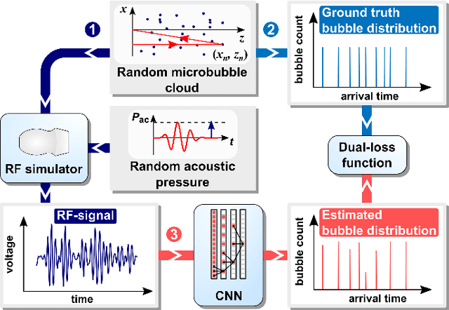
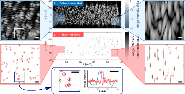
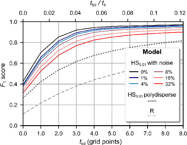
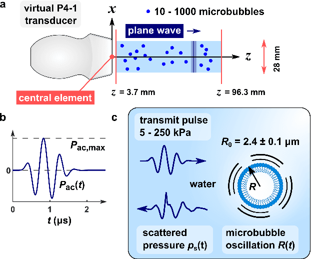
Recently, super-resolution ultrasound imaging with ultrasound localization microscopy (ULM) has received much attention. However, ULM relies on low concentrations of microbubbles in the blood vessels, ultimately resulting in long acquisition times. Here, we present an alternative super-resolution approach, based on direct deconvolution of single-channel ultrasound radio-frequency (RF) signals with a one-dimensional dilated convolutional neural network (CNN). This work focuses on low-frequency ultrasound (1.7 MHz) for deep imaging (10 cm) of a dense cloud of monodisperse microbubbles (up to 1000 microbubbles in the measurement volume, corresponding to an average echo overlap of 94%). Data are generated with a simulator that uses a large range of acoustic pressures (5-250 kPa) and captures the full, nonlinear response of resonant, lipid-coated microbubbles. The network is trained with a novel dual-loss function, which features elements of both a classification loss and a regression loss and improves the detection-localization characteristics of the output. Whereas imposing a localization tolerance of 0 yields poor detection metrics, imposing a localization tolerance corresponding to 4% of the wavelength yields a precision and recall of both 0.90. Furthermore, the detection improves with increasing acoustic pressure and deteriorates with increasing microbubble density. The potential of the presented approach to super-resolution ultrasound imaging is demonstrated with a delay-and-sum reconstruction with deconvolved element data. The resulting image shows an order-of-magnitude gain in axial resolution compared to a delay-and-sum reconstruction with unprocessed element data.