Natalie M. Zahr
Robust thalamic nuclei segmentation from T1-weighted MRI
Apr 14, 2023Abstract:Accurate segmentation of thalamic nuclei, crucial for understanding their role in healthy cognition and in pathologies, is challenging to achieve on standard T1-weighted (T1w) magnetic resonance imaging (MRI) due to poor image contrast. White-matter-nulled (WMn) MRI sequences improve intrathalamic contrast but are not part of clinical protocols or extant databases. Here, we introduce Histogram-based polynomial synthesis (HIPS), a fast preprocessing step that synthesizes WMn-like image contrast from standard T1w MRI using a polynomial approximation. HIPS was incorporated into our Thalamus Optimized Multi-Atlas Segmentation (THOMAS) pipeline, developed and optimized for WMn MRI. HIPS-THOMAS was compared to a convolutional neural network (CNN)-based segmentation method and THOMAS modified for T1w images (T1w-THOMAS). The robustness and accuracy of the three methods were tested across different image contrasts, scanner manufacturers, and field strength. HIPS-synthesized images improved intra-thalamic contrast and thalamic boundaries, and their segmentations yielded significantly better mean Dice, lower percentage of volume error, and lower standard deviations compared to both the CNN method and T1w-THOMAS. Finally, using THOMAS, HIPS-synthesized images were as effective as WMn images for identifying thalamic nuclei atrophy in alcohol use disorders subjects relative to healthy controls, with a higher area under the ROC curve compared to T1w-THOMAS (0.79 vs 0.73).
A Contrast Synthesized Thalamic Nuclei Segmentation Scheme using Convolutional Neural Networks
Dec 17, 2020


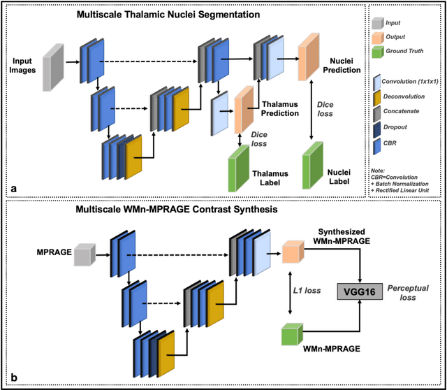
Abstract:Thalamic nuclei have been implicated in several neurological diseases. WMn-MPRAGE images have been shown to provide better intra-thalamic nuclear contrast compared to conventional MPRAGE images but the additional acquisition results in increased examination times. In this work, we investigated 3D Convolutional Neural Network (CNN) based techniques for thalamic nuclei parcellation from conventional MPRAGE images. Two 3D CNNs were developed and compared for thalamic nuclei parcellation using MPRAGE images: a) a native contrast segmentation (NCS) and b) a synthesized contrast segmentation (SCS) using WMn-MPRAGE images synthesized from MPRAGE images. We trained the two segmentation frameworks using MPRAGE images (n=35) and thalamic nuclei labels generated on WMn-MPRAGE images using a multi-atlas based parcellation technique. The segmentation accuracy and clinical utility were evaluated on a cohort comprising of healthy subjects and patients with alcohol use disorder (AUD) (n=45). The SCS network yielded higher Dice scores in the Medial geniculate nucleus (P=.003) and Centromedian nucleus (P=.01) with lower volume differences for Ventral anterior (P=.001) and Ventral posterior lateral (P=.01) nuclei when compared to the NCS network. A Bland-Altman analysis revealed tighter limits of agreement with lower coefficient of variation between true volumes and those predicted by the SCS network. The SCS network demonstrated a significant atrophy in Ventral lateral posterior nucleus in AUD patients compared to healthy age-matched controls (P=0.01), agreeing with previous studies on thalamic atrophy in alcoholism, whereas the NCS network showed spurious atrophy of the Ventral posterior lateral nucleus. CNN-based contrast synthesis prior to segmentation can provide fast and accurate thalamic nuclei segmentation from conventional MPRAGE images.
 Add to Chrome
 Add to Firefox
 Add to Edge