Narges Saeedizadeh
COVID CT-Net: Predicting Covid-19 From Chest CT Images Using Attentional Convolutional Network
Sep 10, 2020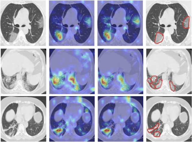
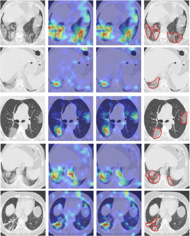
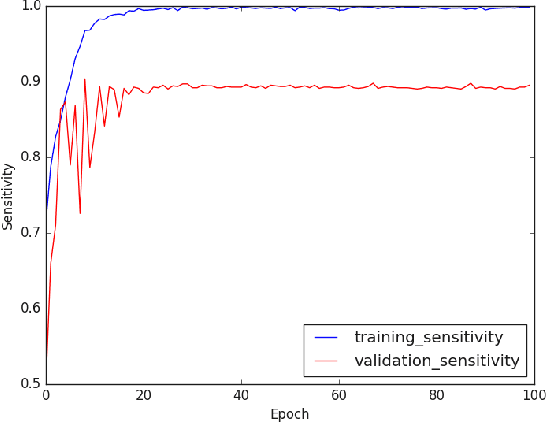
Abstract:The novel corona-virus disease (COVID-19) pandemic has caused a major outbreak in more than 200 countries around the world, leading to a severe impact on the health and life of many people globally. As of Aug 25th of 2020, more than 20 million people are infected, and more than 800,000 death are reported. Computed Tomography (CT) images can be used as a as an alternative to the time-consuming "reverse transcription polymerase chain reaction (RT-PCR)" test, to detect COVID-19. In this work we developed a deep learning framework to predict COVID-19 from CT images. We propose to use an attentional convolution network, which can focus on the infected areas of chest, enabling it to perform a more accurate prediction. We trained our model on a dataset of more than 2000 CT images, and report its performance in terms of various popular metrics, such as sensitivity, specificity, area under the curve, and also precision-recall curve, and achieve very promising results. We also provide a visualization of the attention maps of the model for several test images, and show that our model is attending to the infected regions as intended. In addition to developing a machine learning modeling framework, we also provide the manual annotation of the potentionally infected regions of chest, with the help of a board-certified radiologist, and make that publicly available for other researchers.
COVID TV-UNet: Segmenting COVID-19 Chest CT Images Using Connectivity Imposed U-Net
Aug 06, 2020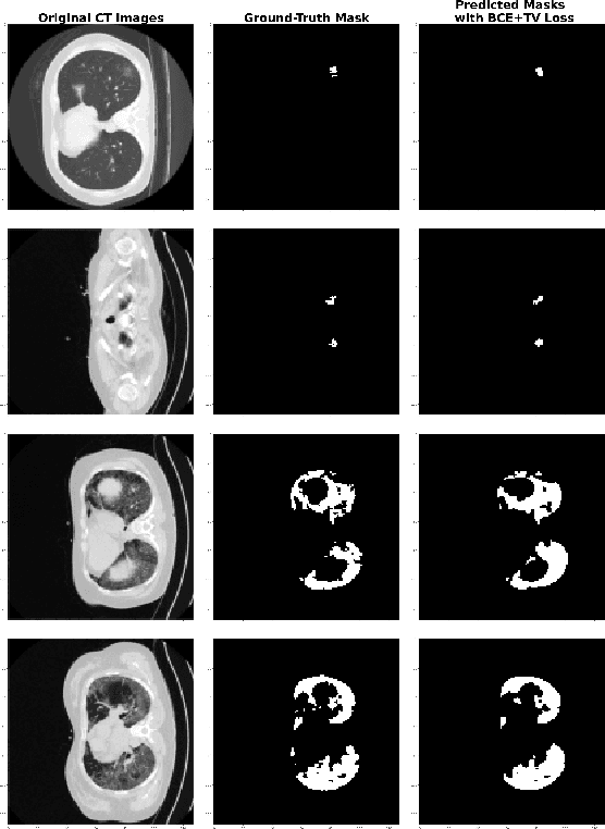
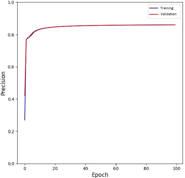
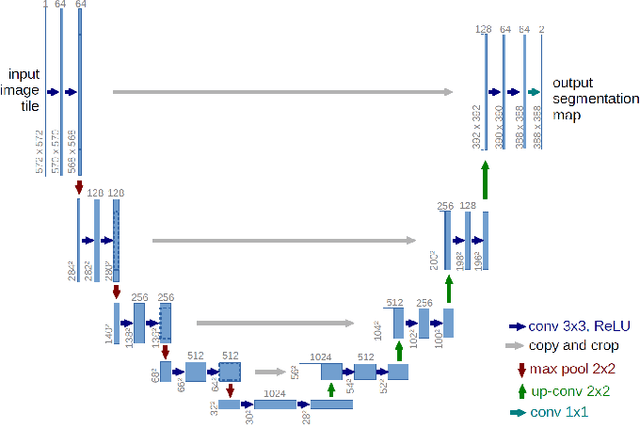
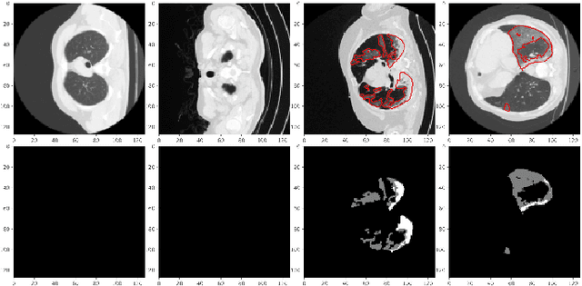
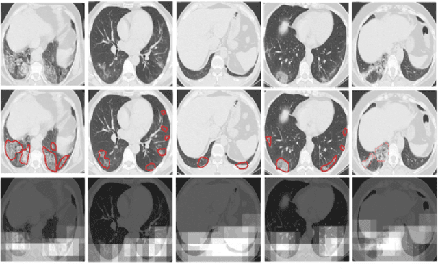
Abstract:The novel corona-virus disease (COVID-19) pandemic has caused a major outbreak in more than 200 countries around the world, leading to a severe impact on the health and life of many people globally. As of mid-July 2020, more than 12 million people were infected, and more than 570,000 death were reported. Computed Tomography (CT) images can be used as an alternative to the time-consuming RT-PCR test, to detect COVID-19. In this work we propose a segmentation framework to detect chest regions in CT images, which are infected by COVID-19. We use an architecture similar to U-Net model, and train it to detect ground glass regions, on pixel level. As the infected regions tend to form a connected component (rather than randomly distributed pixels), we add a suitable regularization term to the loss function, to promote connectivity of the segmentation map for COVID-19 pixels. 2D-anisotropic total-variation is used for this purpose, and therefore the proposed model is called "TV-UNet". Through experimental results on a relatively large-scale CT segmentation dataset of around 900 images, we show that adding this new regularization term leads to 2\% gain on overall segmentation performance compared to the U-Net model. Our experimental analysis, ranging from visual evaluation of the predicted segmentation results to quantitative assessment of segmentation performance (precision, recall, Dice score, and mIoU) demonstrated great ability to identify COVID-19 associated regions of the lungs, achieving a mIoU rate of over 99\%, and a Dice score of around 86\%.
 Add to Chrome
 Add to Firefox
 Add to Edge