Hongbin Tu
Curvilinear object segmentation in medical images based on ODoS filter and deep learning network
Jan 18, 2023Abstract:Automatic segmentation of curvilinear objects in medical images plays an important role in the diagnosis and evaluation of human diseases, yet it is a challenging uncertainty for the complex segmentation task due to different issues like various image appearance, low contrast between curvilinear objects and their surrounding backgrounds, thin and uneven curvilinear structures, and improper background illumination. To overcome these challenges, we present a unique curvilinear structure segmentation framework based on oriented derivative of stick (ODoS) filter and deep learning network for curvilinear object segmentation in medical images. Currently, a large number of deep learning models emphasis on developing deep architectures and ignore capturing the structural features of curvature objects, which may lead to unsatisfactory results. In consequence, a new approach that incorporates the ODoS filter as part of a deep learning network is presented to improve the spatial attention of curvilinear objects. In which, the original image is considered as principal part to describe various image appearance and complex background illumination, the multi-step strategy is used to enhance contrast between curvilinear objects and their surrounding backgrounds, and the vector field is applied to discriminate thin and uneven curvilinear structures. Subsequently, a deep learning framework is employed to extract varvious structural features for curvilinear object segmentation in medical images. The performance of the computational model was validated in experiments with publicly available DRIVE, STARE and CHASEDB1 datasets. Experimental results indicate that the presented model has yielded surprising results compared with some state-of-the-art methods.
Pulmonary Fissure Segmentation in CT Images Based on ODoS Filter and Shape Features
Jan 23, 2022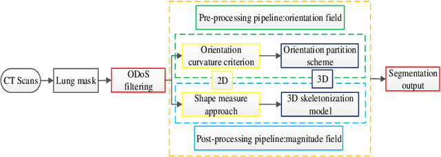
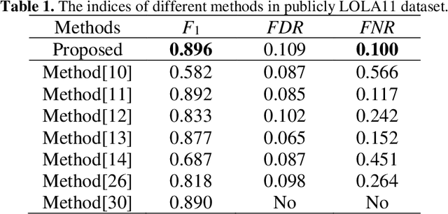
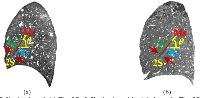

Abstract:Priori knowledge of pulmonary anatomy plays a vital role in diagnosis of lung diseases. In CT images, pulmonary fissure segmentation is a formidable mission due to various of factors. To address the challenge, an useful approach based on ODoS filter and shape features is presented for pulmonary fissure segmentation. Here, we adopt an ODoS filter by merging the orientation information and magnitude information to highlight structure features for fissure enhancement, which can effectively distinguish between pulmonary fissures and clutters. Motivated by the fact that pulmonary fissures appear as linear structures in 2D space and planar structures in 3D space in orientation field, an orientation curvature criterion and an orientation partition scheme are fused to separate fissure patches and other structures in different orientation partition, which can suppress parts of clutters. Considering the shape difference between pulmonary fissures and tubular structures in magnitude field, a shape measure approach and a 3D skeletonization model are combined to segment pulmonary fissures for clutters removal. When applying our scheme to 55 chest CT scans which acquired from a publicly available LOLA11 datasets, the median F1-score, False Discovery Rate (FDR), and False Negative Rate (FNR) respectively are 0.896, 0.109, and 0.100, which indicates that the presented method has a satisfactory pulmonary fissure segmentation performance.
Automatic segmentation of novel coronavirus pneumonia lesions in CT images utilizing deep-supervised ensemble learning network
Nov 17, 2021
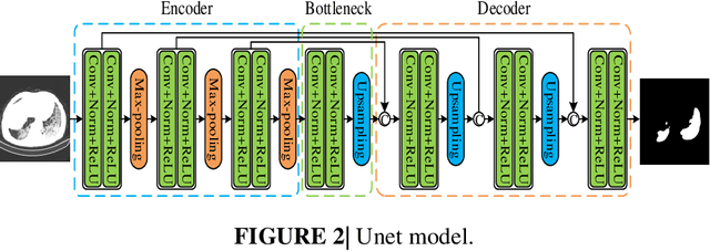

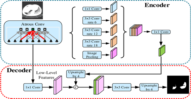
Abstract:Background: The 2019 novel coronavirus disease (COVID-19) has been spread widely in the world, causing a huge threat to people's living environment. Objective: Under computed tomography (CT) imaging, the structure features of COVID-19 lesions are complicated and varied greatly in different cases. To accurately locate COVID-19 lesions and assist doctors to make the best diagnosis and treatment plan, a deep-supervised ensemble learning network is presented for COVID-19 lesion segmentation in CT images. Methods: Considering the fact that a large number of COVID-19 CT images and the corresponding lesion annotations are difficult to obtained, a transfer learning strategy is employed to make up for the shortcoming and alleviate the overfitting problem. Based on the reality that traditional single deep learning framework is difficult to extract COVID-19 lesion features effectively, which may cause some lesions to be undetected. To overcome the problem, a deep-supervised ensemble learning network is presented to combine with local and global features for COVID-19 lesion segmentation. Results: The performance of the proposed method was validated in experiments with a publicly available dataset. Compared with manual annotations, the proposed method acquired a high intersection over union (IoU) of 0.7279. Conclusion: A deep-supervised ensemble learning network was presented for coronavirus pneumonia lesion segmentation in CT images. The effectiveness of the proposed method was verified by visual inspection and quantitative evaluation. Experimental results shown that the proposed mehtod has a perfect performance in COVID-19 lesion segmentation.
 Add to Chrome
 Add to Firefox
 Add to Edge