Hervé Rigneault
Interferometric single-pixel imaging with a multicore fiber
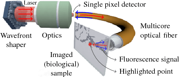
Jul 17, 2023Abstract:Lensless illumination single-pixel imaging with a multicore fiber (MCF) is a computational imaging technique that enables potential endoscopic observations of biological samples at cellular scale. In this work, we show that this technique is tantamount to collecting multiple symmetric rank-one projections (SROP) of a Hermitian \emph{interferometric} matrix -- a matrix encoding the spectral content of the sample image. In this model, each SROP is induced by the complex \emph{sketching} vector shaping the incident light wavefront with a spatial light modulator (SLM), while the projected interferometric matrix collects up to $O(Q^2)$ image frequencies for a $Q$-core MCF. While this scheme subsumes previous sensing modalities, such as raster scanning (RS) imaging with beamformed illumination, we demonstrate that collecting the measurements of $M$ random SLM configurations -- and thus acquiring $M$ SROPs -- allows us to estimate an image of interest if $M$ and $Q$ scale linearly (up to log factors) with the image sparsity level, hence requiring much fewer observations than RS imaging or a complete Nyquist sampling of the $Q \times Q$ interferometric matrix. This demonstration is achieved both theoretically, with a specific restricted isometry analysis of the sensing scheme, and with extensive Monte Carlo experiments. Experimental results made on an actual MCF system finally demonstrate the effectiveness of this imaging procedure on a benchmark image.
Interferometric lensless imaging: rank-one projections of image frequencies with speckle illuminations
Jun 29, 2023Abstract:Lensless illumination single-pixel imaging with a multicore fiber (MCF) is a computational imaging technique that enables potential endoscopic observations of biological samples at cellular scale. In this work, we show that this technique is tantamount to collecting multiple symmetric rank-one projections (SROP) of an interferometric matrix--a matrix encoding the spectral content of the sample image. In this model, each SROP is induced by the complex sketching vector shaping the incident light wavefront with a spatial light modulator (SLM), while the projected interferometric matrix collects up to $O(Q^2)$ image frequencies for a $Q$-core MCF. While this scheme subsumes previous sensing modalities, such as raster scanning (RS) imaging with beamformed illumination, we demonstrate that collecting the measurements of $M$ random SLM configurations--and thus acquiring $M$ SROPs--allows us to estimate an image of interest if $M$ and $Q$ scale log-linearly with the image sparsity level This demonstration is achieved both theoretically, with a specific restricted isometry analysis of the sensing scheme, and with extensive Monte Carlo experiments. On a practical side, we perform a single calibration of the sensing system robust to certain deviations to the theoretical model and independent of the sketching vectors used during the imaging phase. Experimental results made on an actual MCF system demonstrate the effectiveness of this imaging procedure on a benchmark image.
Compressive lensless endoscopy with partial speckle scanning
Apr 22, 2021


Abstract:The lensless endoscope (LE) is a promising device to acquire in vivo images at a cellular scale. The tiny size of the probe enables a deep exploration of the tissues. Lensless endoscopy with a multicore fiber (MCF) commonly uses a spatial light modulator (SLM) to coherently combine, at the output of the MCF, few hundreds of beamlets into a focus spot. This spot is subsequently scanned across the sample to generate a fluorescent image. We propose here a novel scanning scheme, partial speckle scanning (PSS), inspired by compressive sensing theory, that avoids the use of an SLM to perform fluorescent imaging in LE with reduced acquisition time. Such a strategy avoids photo-bleaching while keeping high reconstruction quality. We develop our approach on two key properties of the LE: (i) the ability to easily generate speckles, and (ii) the memory effect in MCF that allows to use fast scan mirrors to shift light patterns. First, we show that speckles are sub-exponential random fields. Despite their granular structure, an appropriate choice of the reconstruction parameters makes them good candidates to build efficient sensing matrices. Then, we numerically validate our approach and apply it on experimental data. The proposed sensing technique outperforms conventional raster scanning: higher reconstruction quality is achieved with far fewer observations. For a fixed reconstruction quality, our speckle scanning approach is faster than compressive sensing schemes which require to change the speckle pattern for each observation.
Compressive Sampling Approach for Image Acquisition with Lensless Endoscope
Oct 30, 2018


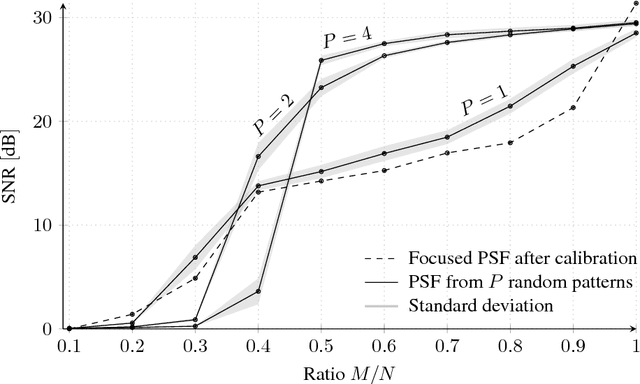
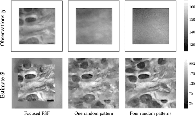
Abstract:The lensless endoscope is a promising device designed to image tissues in vivo at the cellular scale. The traditional acquisition setup consists in raster scanning during which the focused light beam from the optical fiber illuminates sequentially each pixel of the field of view (FOV). The calibration step to focus the beam and the sampling scheme both take time. In this preliminary work, we propose a scanning method based on compressive sampling theory. The method does not rely on a focused beam but rather on the random illumination patterns generated by the single-mode fibers. Experiments are performed on synthetic data for different compression rates (from 10 to 100% of the FOV).
 Add to Chrome
 Add to Firefox
 Add to Edge