Helena Mehtonen
Interactive 3D Segmentation for Primary Gross Tumor Volume in Oropharyngeal Cancer
Sep 10, 2024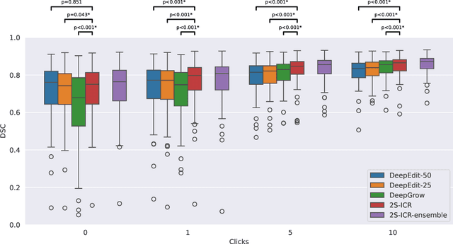


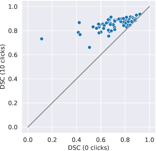
Abstract:The main treatment modality for oropharyngeal cancer (OPC) is radiotherapy, where accurate segmentation of the primary gross tumor volume (GTVp) is essential. However, accurate GTVp segmentation is challenging due to significant interobserver variability and the time-consuming nature of manual annotation, while fully automated methods can occasionally fail. An interactive deep learning (DL) model offers the advantage of automatic high-performance segmentation with the flexibility for user correction when necessary. In this study, we examine interactive DL for GTVp segmentation in OPC. We implement state-of-the-art algorithms and propose a novel two-stage Interactive Click Refinement (2S-ICR) framework. Using the 2021 HEad and neCK TumOR (HECKTOR) dataset for development and an external dataset from The University of Texas MD Anderson Cancer Center for evaluation, the 2S-ICR framework achieves a Dice similarity coefficient of 0.713 $\pm$ 0.152 without user interaction and 0.824 $\pm$ 0.099 after five interactions, outperforming existing methods in both cases.
Comparison of Deep Learning Segmentation and Multigrader-annotated Mandibular Canals of Multicenter CBCT scans
May 27, 2022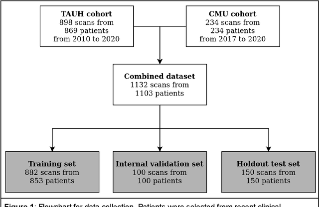
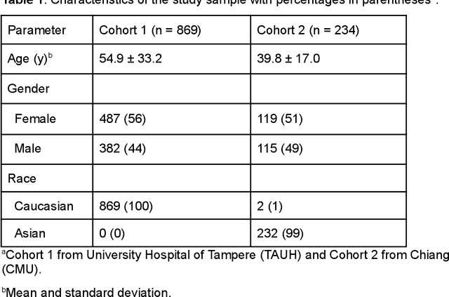
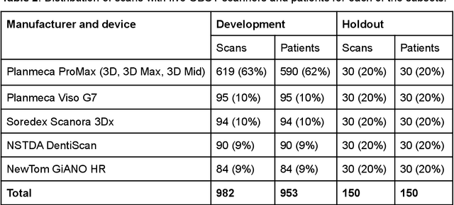
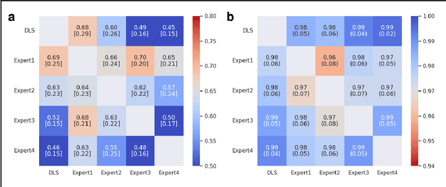
Abstract:Deep learning approach has been demonstrated to automatically segment the bilateral mandibular canals from CBCT scans, yet systematic studies of its clinical and technical validation are scarce. To validate the mandibular canal localization accuracy of a deep learning system (DLS) we trained it with 982 CBCT scans and evaluated using 150 scans of five scanners from clinical workflow patients of European and Southeast Asian Institutes, annotated by four radiologists. The interobserver variability was compared to the variability between the DLS and the radiologists. In addition, the generalization of DLS to CBCT scans from scanners not used in the training data was examined to evaluate the out-of-distribution generalization capability. The DLS had lower variability to the radiologists than the interobserver variability between them and it was able to generalize to three new devices. For the radiologists' consensus segmentation, used as gold standard, the DLS had a symmetric mean curve distance of 0.39 mm compared to those of the individual radiologists with 0.62 mm, 0.55 mm, 0.47 mm, and 0.42 mm. The DLS showed comparable or slightly better performance in the segmentation of the mandibular canal with the radiologists and generalization capability to new scanners.
 Add to Chrome
 Add to Firefox
 Add to Edge