Gayathri Malamal
Towards Non-contact 3D Ultrasound for Wrist Imaging
Oct 06, 2023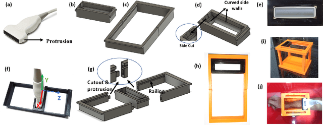

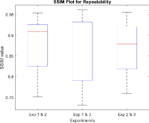
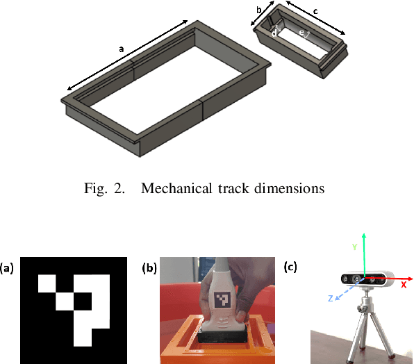
Abstract:Objective: The objective of this work is an attempt towards non-contact freehand 3D ultrasound imaging with minimal complexity added to the existing point of care ultrasound (POCUS) systems. Methods: This study proposes a novel approach of using a mechanical track for non-contact ultrasound (US) scanning. The approach thus restricts the probe motion to a linear plane, to simplify the acquisition and 3D reconstruction process. A pipeline for US 3D volume reconstruction employing an US research platform and a GPU-based edge device is developed. Results: The efficacy of the proposed approach is demonstrated through ex-vivo and in-vivo experiments. Conclusion: The proposed approach with the adjustable field of view capability, non-contact design, and low cost of deployment without significantly altering the existing setup would open doors for up gradation of traditional systems to a wide range of 3D US imaging applications. Significance: Ultrasound (US) imaging is a popular clinical imaging modality for the point-of-care bedside imaging, particularly of the wrist/knee in the pediatric population due to its non-invasive and radiation free nature. However, the limited views of tissue structures obtained with 2D US in such scenarios make the diagnosis challenging. To overcome this, 3D US imaging which uses 2D US images and their orientation/position to reconstruct 3D volumes was developed. The accurate position estimation of the US probe at low cost has always stood as a challenging task in 3D reconstruction. Additionally, US imaging involves contact, which causes difficulty to pediatric subjects while monitoring live fractures or open wounds. Towards overcoming these challenges, a novel framework is attempted in this work.
Fast Marching based Tissue Adaptive Delay Estimation for Aberration Corrected Delay and Sum Beamforming in Ultrasound Imaging
Apr 19, 2023Abstract:Conventional ultrasound (US) imaging employs the delay and sum (DAS) receive beamforming with dynamic receive focus for image reconstruction due to its simplicity and robustness. However, the DAS beamforming follows a geometrical method of delay estimation with a spatially constant speed-of-sound (SoS) of 1540 m/s throughout the medium irrespective of the tissue in-homogeneity. This approximation leads to errors in delay estimations that accumulate with depth and degrades the resolution, contrast and overall accuracy of the US image. In this work, we propose a fast marching based DAS for focused transmissions which leverages the approximate SoS map to estimate the refraction corrected propagation delays for each pixel in the medium. The proposed approach is validated qualitatively and quantitatively for imaging depths of upto ~ 11 cm through simulations, where fat layer induced aberration is employed to alter the SoS in the medium. To the best of authors' knowledge, this is the first work considering the effect of SoS on image quality for deeper imaging.
A Simplified 3D Ultrasound Freehand Imaging Framework Using 1D Linear Probe and Low-Cost Mechanical Track
Feb 16, 2023Abstract:Ultrasound imaging is the most popular medical imaging modality for point-of-care bedside imaging. However, 2D ultrasound imaging provides only limited views of the organ of interest, making diagnosis challenging. To overcome this, 3D ultrasound imaging was developed, which uses 2D ultrasound images and their orientation/position to reconstruct 3D volumes. The accurate position estimation of the ultrasound probe at low cost has always stood as a challenging task in 3D reconstruction. In this study, we propose a novel approach of using a mechanical track for ultrasound scanning, which restricts the probe motion to a linear plane, simplifying the acquisition and hence the reconstruction process. We also present an end-to-end pipeline for 3D ultrasound volume reconstruction and demonstrate its efficacy with an in-vitro tube phantom study and an ex-vivo bone experiment. The comparison between a sensorless freehand and the proposed mechanical track based acquisition is available online (shorturl.at/jqvX0).
Learning while Acquisition: Towards Active Learning Framework for Beamforming in Ultrasound Imaging
Jul 31, 2022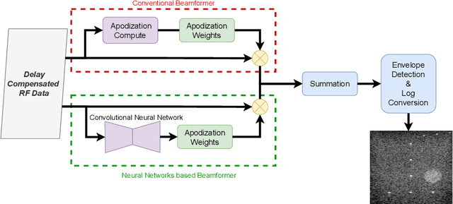

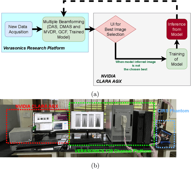

Abstract:In the recent past, there have been many efforts to accelerate adaptive beamforming for ultrasound (US) imaging using neural networks (NNs). However, most of these efforts are based on static models, i.e., they are trained to learn a single adaptive beamforming approach (e.g., minimum variance distortionless response (MVDR)) assuming that they result in the best image quality. Moreover, the training of such NNs is initiated only after acquiring a large set of data that consumes several gigabytes (GBs) of storage space. In this study, an active learning framework for beamforming is described for the first time in the context of NNs. The best quality image chosen by the user serves as the ground truth for the proposed technique, which trains the NN concurrently with data acqusition. On average, the active learning approach takes 0.5 seconds to complete a single iteration of training.
Introducing Introspective Transmission for Reflection Characterization in High Frame-Rate Ultrasound Imaging
Jul 16, 2021
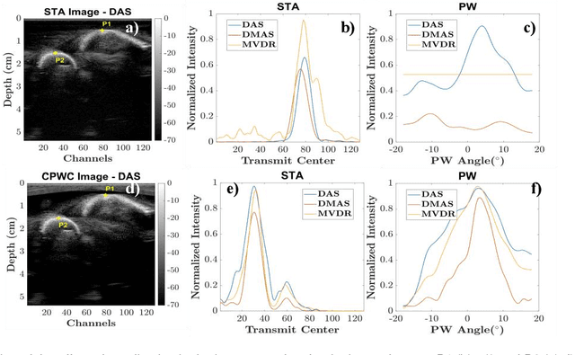
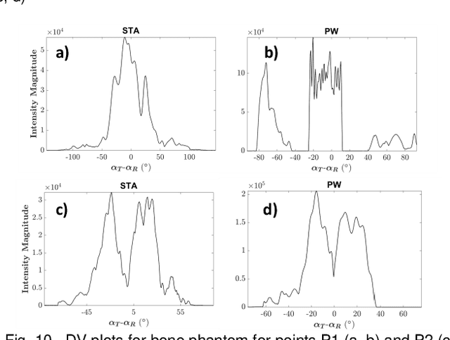
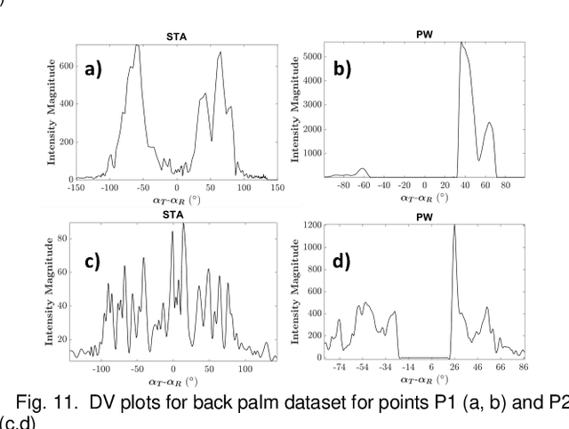
Abstract:In ultrasound imaging, most of the transmit and receive beamforming schemes assume a homogenous diffuse medium and are evaluated based on contrast, temporal and spatial resolutions. However, most medium are constituted by both diffuse and specular regions and the assumption of a homogeneous medium does not hold good in all cases. Eventhough, there are some adaptive beamforming approaches proposed in literature, they are mostly for receive beamforming. This study is aimed at investigating the relevance of transmission schemes in characterizing the diffuse and specular reflections, particularly at high frame rates. The transmit wavefront interaction behavior on the tissue interfaces for two high frame-rate transmission modalities, i.e. conventional synthetic transmit aperture imaging and multi-angle plane-wave imaging, are analyzed for multiple in-vitro and in-vivo radio-frequency datasets. Two novel visualization perspectives are proposed called contour isolines and directivity variance to understand the wave interaction with different tissue interfaces by considering the scalar and vector aspects of the reflected intensities respectively. We also rationalize the relevance of choosing the appropriate receive beamforming scheme according to the transmission modality through a comparison of delay and sum, filtered delay multiply and sum, minimum variance distortionless response, and specular beamforming algorithms. It is found that a synergistic blend of transmit and receive beamforming schemes adaptive to the tissue is inevitable to avoid any misdiagnosis.
 Add to Chrome
 Add to Firefox
 Add to Edge