Evelyn Eger
LIENS, INRIA Paris - Rocquencourt
Multi-scale Mining of fMRI data with Hierarchical Structured Sparsity
Dec 08, 2011

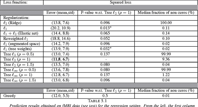

Abstract:Inverse inference, or "brain reading", is a recent paradigm for analyzing functional magnetic resonance imaging (fMRI) data, based on pattern recognition and statistical learning. By predicting some cognitive variables related to brain activation maps, this approach aims at decoding brain activity. Inverse inference takes into account the multivariate information between voxels and is currently the only way to assess how precisely some cognitive information is encoded by the activity of neural populations within the whole brain. However, it relies on a prediction function that is plagued by the curse of dimensionality, since there are far more features than samples, i.e., more voxels than fMRI volumes. To address this problem, different methods have been proposed, such as, among others, univariate feature selection, feature agglomeration and regularization techniques. In this paper, we consider a sparse hierarchical structured regularization. Specifically, the penalization we use is constructed from a tree that is obtained by spatially-constrained agglomerative clustering. This approach encodes the spatial structure of the data at different scales into the regularization, which makes the overall prediction procedure more robust to inter-subject variability. The regularization used induces the selection of spatially coherent predictive brain regions simultaneously at different scales. We test our algorithm on real data acquired to study the mental representation of objects, and we show that the proposed algorithm not only delineates meaningful brain regions but yields as well better prediction accuracy than reference methods.
A supervised clustering approach for fMRI-based inference of brain states
Apr 28, 2011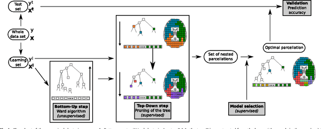

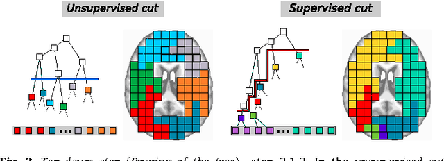

Abstract:We propose a method that combines signals from many brain regions observed in functional Magnetic Resonance Imaging (fMRI) to predict the subject's behavior during a scanning session. Such predictions suffer from the huge number of brain regions sampled on the voxel grid of standard fMRI data sets: the curse of dimensionality. Dimensionality reduction is thus needed, but it is often performed using a univariate feature selection procedure, that handles neither the spatial structure of the images, nor the multivariate nature of the signal. By introducing a hierarchical clustering of the brain volume that incorporates connectivity constraints, we reduce the span of the possible spatial configurations to a single tree of nested regions tailored to the signal. We then prune the tree in a supervised setting, hence the name supervised clustering, in order to extract a parcellation (division of the volume) such that parcel-based signal averages best predict the target information. Dimensionality reduction is thus achieved by feature agglomeration, and the constructed features now provide a multi-scale representation of the signal. Comparisons with reference methods on both simulated and real data show that our approach yields higher prediction accuracy than standard voxel-based approaches. Moreover, the method infers an explicit weighting of the regions involved in the regression or classification task.
Total variation regularization for fMRI-based prediction of behaviour
Feb 05, 2011
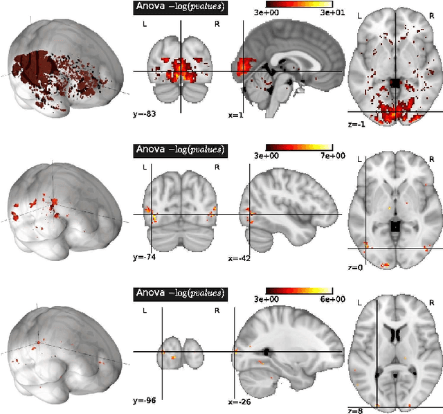
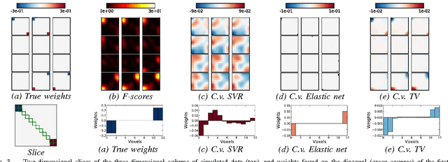
Abstract:While medical imaging typically provides massive amounts of data, the extraction of relevant information for predictive diagnosis remains a difficult challenge. Functional MRI (fMRI) data, that provide an indirect measure of task-related or spontaneous neuronal activity, are classically analyzed in a mass-univariate procedure yielding statistical parametric maps. This analysis framework disregards some important principles of brain organization: population coding, distributed and overlapping representations. Multivariate pattern analysis, i.e., the prediction of behavioural variables from brain activation patterns better captures this structure. To cope with the high dimensionality of the data, the learning method has to be regularized. However, the spatial structure of the image is not taken into account in standard regularization methods, so that the extracted features are often hard to interpret. More informative and interpretable results can be obtained with the l_1 norm of the image gradient, a.k.a. its Total Variation (TV), as regularization. We apply for the first time this method to fMRI data, and show that TV regularization is well suited to the purpose of brain mapping while being a powerful tool for brain decoding. Moreover, this article presents the first use of TV regularization for classification.
 Add to Chrome
 Add to Firefox
 Add to Edge