Eduardo V. Navajas
Department of Ophthalmology and Visual Sciences, University of British Columbia, Canada
Microvasculature Segmentation and Inter-capillary Area Quantification of the Deep Vascular Complex using Transfer Learning
Mar 19, 2020


Abstract:Purpose: Optical Coherence Tomography Angiography (OCT-A) permits visualization of the changes to the retinal circulation due to diabetic retinopathy (DR), a microvascular complication of diabetes. We demonstrate accurate segmentation of the vascular morphology for the superficial capillary plexus and deep vascular complex (SCP and DVC) using a convolutional neural network (CNN) for quantitative analysis. Methods: Retinal OCT-A with a 6x6mm field of view (FOV) were acquired using a Zeiss PlexElite. Multiple-volume acquisition and averaging enhanced the vessel network contrast used for training the CNN. We used transfer learning from a CNN trained on 76 images from smaller FOVs of the SCP acquired using different OCT systems. Quantitative analysis of perfusion was performed on the automated vessel segmentations in representative patients with DR. Results: The automated segmentations of the OCT-A images maintained the hierarchical branching and lobular morphologies of the SCP and DVC, respectively. The network segmented the SCP with an accuracy of 0.8599, and a Dice index of 0.8618. For the DVC, the accuracy was 0.7986, and the Dice index was 0.8139. The inter-rater comparisons for the SCP had an accuracy and Dice index of 0.8300 and 0.6700, respectively, and 0.6874 and 0.7416 for the DVC. Conclusions: Transfer learning reduces the amount of manually-annotated images required, while producing high quality automatic segmentations of the SCP and DVC. Using high quality training data preserves the characteristic appearance of the capillary networks in each layer. Translational Relevance: Accurate retinal microvasculature segmentation with the CNN results in improved perfusion analysis in diabetic retinopathy.
Deep learning vessel segmentation and quantification of the foveal avascular zone using commercial and prototype OCT-A platforms
Sep 25, 2019


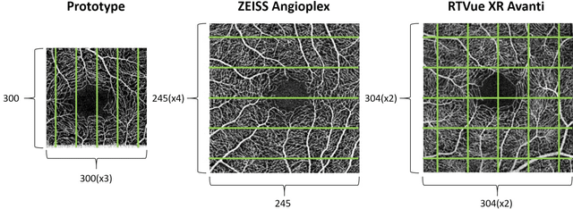
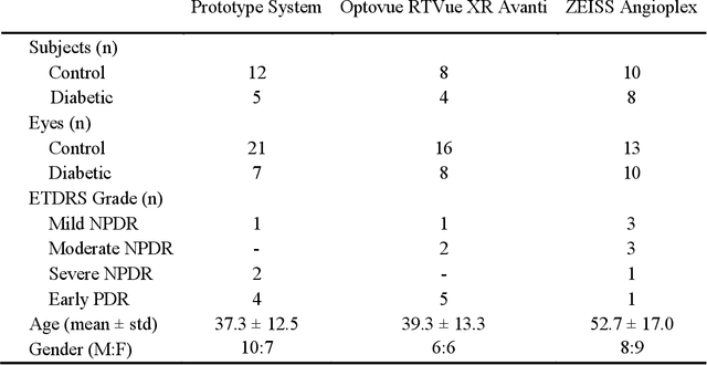
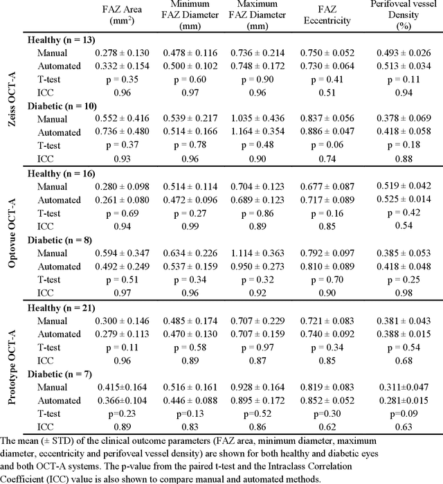
Abstract:Automatic quantification of perifoveal vessel densities in optical coherence tomography angiography (OCT-A) images face challenges such as variable intra- and inter-image signal to noise ratios, projection artefacts from outer vasculature layers, and motion artefacts. This study demonstrates the utility of deep neural networks for automatic quantification of foveal avascular zone (FAZ) parameters and perifoveal vessel density of OCT-A images in healthy and diabetic eyes. OCT-A images of the foveal region were acquired using three OCT-A systems: a 1060nm Swept Source (SS)-OCT prototype, RTVue XR Avanti (Optovue Inc., Fremont, CA), and the ZEISS Angioplex (Carl Zeiss Meditec, Dublin, CA). Automated segmentation was then performed using a deep neural network. Four FAZ morphometric parameters (area, min/max diameter, and eccentricity) and perifoveal vessel density were used as outcome measures. The accuracy, sensitivity and specificity of the DNN vessel segmentations were comparable across all three device platforms. No significant difference between the means of the measurements from automated and manual segmentations were found for any of the outcome measures on any system. The intraclass correlation coefficient (ICC) was also good (> 0.51) for all measurements. Automated deep learning vessel segmentation of OCT-A may be suitable for both commercial and research purposes for better quantification of the retinal circulation.
 Add to Chrome
 Add to Firefox
 Add to Edge