Chin Kuo
Detecting Pulmonary Embolism from Computed Tomography Using Convolutional Neural Network
Jun 03, 2022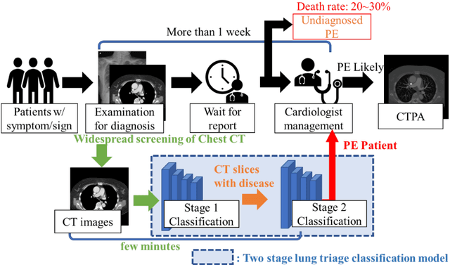


Abstract:The clinical symptoms of pulmonary embolism (PE) are very diverse and non-specific, which makes it difficult to diagnose. In addition, pulmonary embolism has multiple triggers and is one of the major causes of vascular death. Therefore, if it can be detected and treated quickly, it can significantly reduce the risk of death in hospitalized patients. In the detection process, the cost of computed tomography pulmonary angiography (CTPA) is high, and angiography requires the injection of contrast agents, which increase the risk of damage to the patient. Therefore, this study will use a deep learning approach to detect pulmonary embolism in all patients who take a CT image of the chest using a convolutional neural network. With the proposed pulmonary embolism detection system, we can detect the possibility of pulmonary embolism at the same time as the patient's first CT image, and schedule the CTPA test immediately, saving more than a week of CT image screening time and providing timely diagnosis and treatment to the patient.
Computerized Tomography Pulmonary Angiography Image Simulation using Cycle Generative Adversarial Network from Chest CT imaging in Pulmonary Embolism Patients
May 17, 2022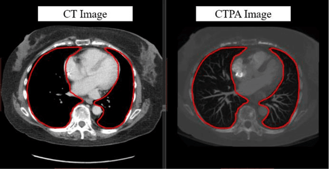
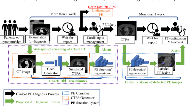
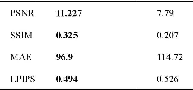
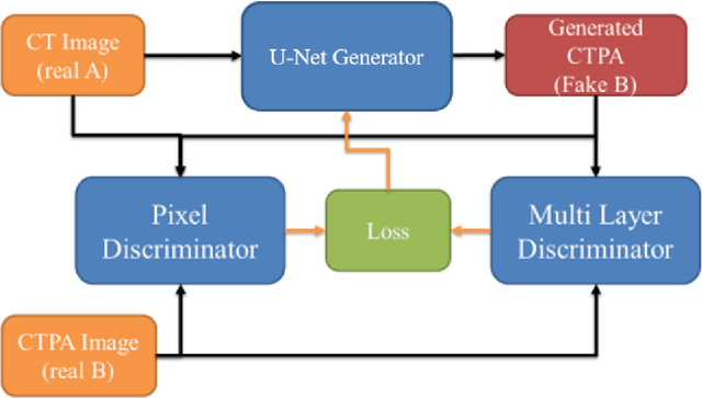
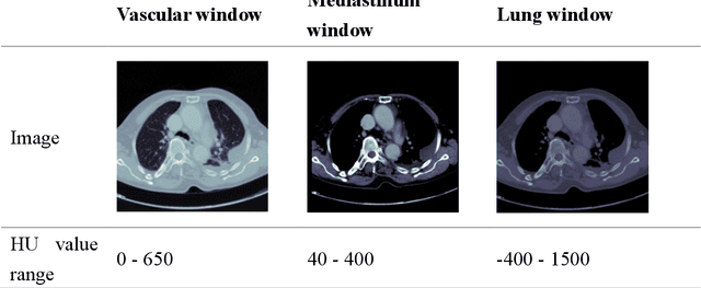
Abstract:The purpose of this research is to develop a system that generates simulated computed tomography pulmonary angiography (CTPA) images clinically for pulmonary embolism diagnoses. Nowadays, CTPA images are the gold standard computerized detection method to determine and identify the symptoms of pulmonary embolism (PE), although performing CTPA is harmful for patients and also expensive. Therefore, we aim to detect possible PE patients through CT images. The system will simulate CTPA images with deep learning models for the identification of PE patients' symptoms, providing physicians with another reference for determining PE patients. In this study, the simulated CTPA image generation system uses a generative antagonistic network to enhance the features of pulmonary vessels in the CT images to strengthen the reference value of the images and provide a basis for hospitals to judge PE patients. We used the CT images of 22 patients from National Cheng Kung University Hospital and the corresponding CTPA images as the training data for the task of simulating CTPA images and generated them using two sets of generative countermeasure networks. This study is expected to propose a new approach to the clinical diagnosis of pulmonary embolism, in which a deep learning network is used to assist in the complex screening process and to review the generated simulated CTPA images, allowing physicians to assess whether a patient needs to undergo detailed testing for CTPA, improving the speed of detection of pulmonary embolism and significantly reducing the number of undetected patients.
Feature-enhanced Adversarial Semi-supervised Semantic Segmentation Network for Pulmonary Embolism Annotation
Apr 08, 2022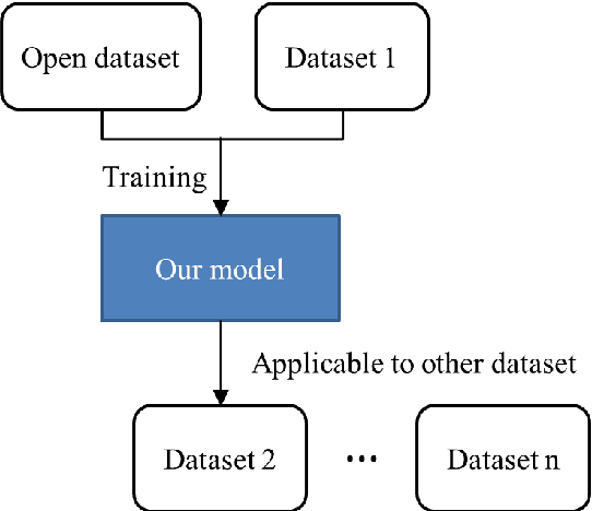

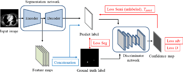
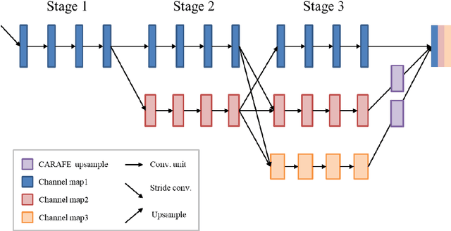
Abstract:This study established a feature-enhanced adversarial semi-supervised semantic segmentation model to automatically annotate pulmonary embolism lesion areas in computed tomography pulmonary angiogram (CTPA) images. In current studies, all of the PE CTPA image segmentation methods are trained by supervised learning. However, the supervised learning models need to be retrained and the images need to be relabeled when the CTPA images come from different hospitals. This study proposed a semi-supervised learning method to make the model applicable to different datasets by adding a small amount of unlabeled images. By training the model with both labeled and unlabeled images, the accuracy of unlabeled images can be improved and the labeling cost can be reduced. Our semi-supervised segmentation model includes a segmentation network and a discriminator network. We added feature information generated from the encoder of segmentation network to the discriminator so that it can learn the similarity between predicted mask and ground truth mask. This HRNet-based architecture can maintain a higher resolution for convolutional operations so the prediction of small PE lesion areas can be improved. We used the labeled open-source dataset and the unlabeled National Cheng Kung University Hospital (NCKUH) (IRB number: B-ER-108-380) dataset to train the semi-supervised learning model, and the resulting mean intersection over union (mIOU), dice score, and sensitivity achieved 0.3510, 0.4854, and 0.4253, respectively on the NCKUH dataset. Then, we fine-tuned and tested the model with a small amount of unlabeled PE CTPA images from China Medical University Hospital (CMUH) (IRB number: CMUH110-REC3-173) dataset. Comparing the results of our semi-supervised model with the supervised model, the mIOU, dice score, and sensitivity improved from 0.2344, 0.3325, and 0.3151 to 0.3721, 0.5113, and 0.4967, respectively.
Convolutional Neural Network for Early Pulmonary Embolism Detection via Computed Tomography Pulmonary Angiography
Apr 07, 2022

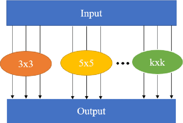
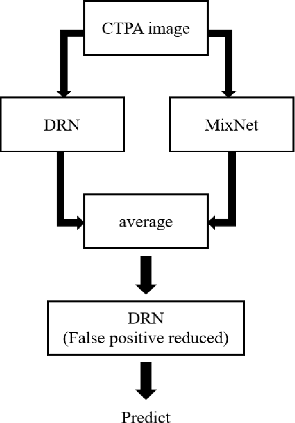
Abstract:This study was conducted to develop a computer-aided detection (CAD) system for triaging patients with pulmonary embolism (PE). The purpose of the system was to reduce the death rate during the waiting period. Computed tomography pulmonary angiography (CTPA) is used for PE diagnosis. Because CTPA reports require a radiologist to review the case and suggest further management, this creates a waiting period during which patients may die. Our proposed CAD method was thus designed to triage patients with PE from those without PE. In contrast to related studies involving CAD systems that identify key PE lesion images to expedite PE diagnosis, our system comprises a novel classification-model ensemble for PE detection and a segmentation model for PE lesion labeling. The models were trained using data from National Cheng Kung University Hospital and open resources. The classification model yielded 0.73 for receiver operating characteristic curve (accuracy = 0.85), while the mean intersection over union was 0.689 for the segmentation model. The proposed CAD system can distinguish between patients with and without PE and automatically label PE lesions to expedite PE diagnosis
 Add to Chrome
 Add to Firefox
 Add to Edge