Augusto Antunes
Predição da Idade Cerebral a partir de Imagens de Ressonância Magnética utilizando Redes Neurais Convolucionais
Dec 23, 2021Abstract:In this work, deep learning techniques for brain age prediction from magnetic resonance images are investigated, aiming to assist in the identification of biomarkers of the natural aging process. The identification of biomarkers is useful for detecting an early-stage neurodegenerative process, as well as for predicting age-related or non-age-related cognitive decline. Two techniques are implemented and compared in this work: a 3D Convolutional Neural Network applied to the volumetric image and a 2D Convolutional Neural Network applied to slices from the axial plane, with subsequent fusion of individual predictions. The best result was obtained by the 2D model, which achieved a mean absolute error of 3.83 years. -- Neste trabalho s\~ao investigadas t\'ecnicas de aprendizado profundo para a predi\c{c}\~ao da idade cerebral a partir de imagens de resson\^ancia magn\'etica, visando auxiliar na identifica\c{c}\~ao de biomarcadores do processo natural de envelhecimento. A identifica\c{c}\~ao de biomarcadores \'e \'util para a detec\c{c}\~ao de um processo neurodegenerativo em est\'agio inicial, al\'em de possibilitar prever um decl\'inio cognitivo relacionado ou n\~ao \`a idade. Duas t\'ecnicas s\~ao implementadas e comparadas neste trabalho: uma Rede Neural Convolucional 3D aplicada na imagem volum\'etrica e uma Rede Neural Convolucional 2D aplicada a fatias do plano axial, com posterior fus\~ao das predi\c{c}\~oes individuais. O melhor resultado foi obtido pelo modelo 2D, que alcan\c{c}ou um erro m\'edio absoluto de 3.83 anos.
Explainable Deep CNNs for MRI-Based Diagnosis of Alzheimer's Disease
Apr 25, 2020


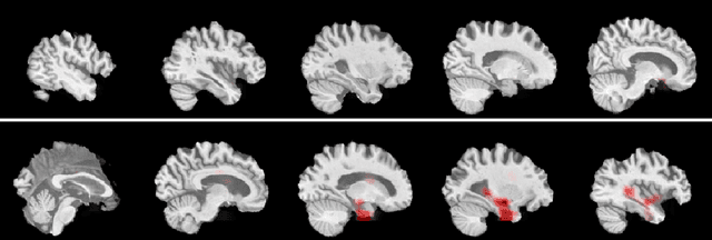
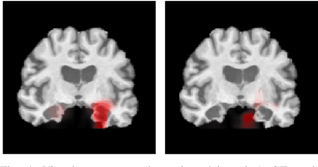
Abstract:Deep Convolutional Neural Networks (CNNs) are becoming prominent models for semi-automated diagnosis of Alzheimer's Disease (AD) using brain Magnetic Resonance Imaging (MRI). Although being highly accurate, deep CNN models lack transparency and interpretability, precluding adequate clinical reasoning and not complying with most current regulatory demands. One popular choice for explaining deep image models is occluding regions of the image to isolate their influence on the prediction. However, existing methods for occluding patches of brain scans generate images outside the distribution to which the model was trained for, thus leading to unreliable explanations. In this paper, we propose an alternative explanation method that is specifically designed for the brain scan task. Our method, which we refer to as Swap Test, produces heatmaps that depict the areas of the brain that are most indicative of AD, providing interpretability for the model's decisions in a format understandable to clinicians. Experimental results using an axiomatic evaluation show that the proposed method is more suitable for explaining the diagnosis of AD using MRI while the opposite trend was observed when using a typical occlusion test. Therefore, we believe our method may address the inherent black-box nature of deep neural networks that are capable of diagnosing AD.
 Add to Chrome
 Add to Firefox
 Add to Edge