Angela Di Fulvio
Deep-learning Segmentation of Small Volumes in CT images for Radiotherapy Treatment Planning
Apr 05, 2024


Abstract:Our understanding of organs at risk is progressing to include physical small tissues such as coronary arteries and the radiosensitivities of many small organs and tissues are high. Therefore, the accurate segmentation of small volumes in external radiotherapy is crucial to protect them from over-irradiation. Moreover, with the development of the particle therapy and on-board imaging, the treatment becomes more accurate and precise. The purpose of this work is to optimize organ segmentation algorithms for small organs. We used 50 three-dimensional (3-D) computed tomography (CT) head and neck images from StructSeg2019 challenge to develop a general-purpose V-Net model to segment 20 organs in the head and neck region. We applied specific strategies to improve the segmentation accuracy of the small volumes in this anatomical region, i.e., the lens of the eye. Then, we used 17 additional head images from OSF healthcare to validate the robustness of the V Net model optimized for small-volume segmentation. With the study of the StructSeg2019 images, we found that the optimization of the image normalization range and classification threshold yielded a segmentation improvement of the lens of the eye of approximately 50%, compared to the use of the V-Net not optimized for small volumes. We used the optimized model to segment 17 images acquired using heterogeneous protocols. We obtained comparable Dice coefficient values for the clinical and StructSeg2019 images (0.61 plus/minus 0.07 and 0.58 plus/minus 0.10 for the left and right lens of the eye, respectively)
Statistical modelling and Bayesian inversion for a Compton imaging system: application to radioactive source localisation
Feb 16, 2024Abstract:This paper presents a statistical forward model for a Compton imaging system, called Compton imager. This system, under development at the University of Illinois Urbana Champaign, is a variant of Compton cameras with a single type of sensors which can simultaneously act as scatterers and absorbers. This imager is convenient for imaging situations requiring a wide field of view. The proposed statistical forward model is then used to solve the inverse problem of estimating the location and energy of point-like sources from observed data. This inverse problem is formulated and solved in a Bayesian framework by using a Metropolis within Gibbs algorithm for the estimation of the location, and an expectation-maximization algorithm for the estimation of the energy. This approach leads to more accurate estimation when compared with the deterministic standard back-projection approach, with the additional benefit of uncertainty quantification in the low photon imaging setting.
Algorithms for TRISO Fuel Identification Based on X-ray CT Validated on Tungsten-Carbide Compacts
Apr 28, 2022


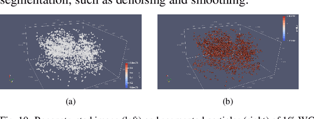
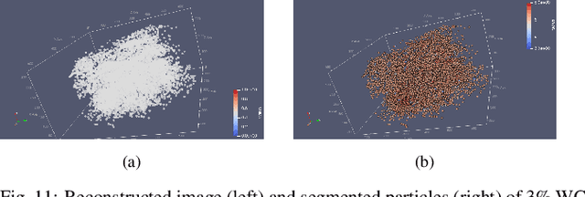
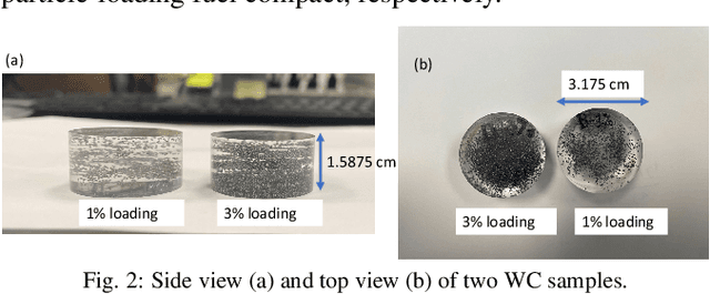
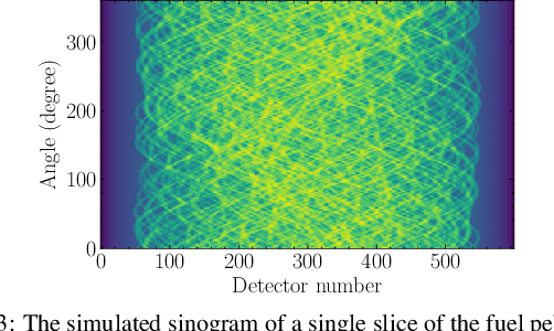
Abstract:Tristructural-isotropic (TRISO) fuel is one of the most mature fuel types for candidate advanced reactor types under development. TRISO-fuel pebbles flow continuously through the reactor core and can be reinserted into the reactor several times until a target burnup is reached. The capability of identifying individual fuel pebbles would allow us to calculate the fuel residence time in the core and validate pebble flow computational models, prevent excessive burnup accumulation or premature fuel discharge, and maintain accountability of special nuclear materials during fuel circulation. In this work, we have developed a 3D image reconstruction and segmentation algorithm to accurately segment TRISO particles and extract the unique 3D distribution. We have developed a rotation-invariant and noise-robust identification algorithm that allows us to identify the pebble and retrieve the pebble ID in the presence of rotations and noises. We also report the results of 200kV X-ray CT image reconstruction of a mock-up fuel sample consisting of tungsten-carbide (WC) kernels in a lucite matrix. The 3D distribution of TRISO particles along with other signatures such as $^{235}$U enrichment and burnup level extracted through neutron multiplicity counting, would enable accurate fuel identification in a reasonable amount of time.
 Add to Chrome
 Add to Firefox
 Add to Edge