 Add to Chrome
 Add to Firefox
 Add to Edge
Perfusion Imaging and Single Material Reconstruction in Polychromatic Photon Counting CT
Feb 02, 2026Background: Perfusion computed tomography (CT) images the dynamics of a contrast agent through the body over time, and is one of the highest X-ray dose scans in medical imaging. Recently, a theoretically justified reconstruction algorithm based on a monotone variational inequality (VI) was proposed for single material polychromatic photon-counting CT, and showed promising early results at low-dose imaging. Purpose: We adapt this reconstruction algorithm for perfusion CT, to reconstruct the concentration map of the contrast agent while the static background tissue is assumed known; we call our method VI-PRISM (VI-based PeRfusion Imaging and Single Material reconstruction). We evaluate its potential for dose-reduced perfusion CT, using a digital phantom with water and iodine of varying concentration. Methods: Simulated iodine concentrations range from 0.05 to 2.5 mg/ml. The simulated X-ray source emits photons up to 100 keV, with average intensity ranging from $10^5$ down to $10^2$ photons per detector element. The number of tomographic projections was varied from 984 down to 8 to characterize the tradeoff in photon allocation between views and intensity. Results: We compare VI-PRISM against filtered back-projection (FBP), and find that VI-PRISM recovers iodine concentration with error below 0.4 mg/ml at all source intensity levels tested. Even with a dose reduction between 10x and 100x compared to FBP, VI-PRISM exhibits reconstruction quality on par with FBP. Conclusion: Across all photon budgets and angular sampling densities tested, VI-PRISM achieved consistently lower RMSE, reduced noise, and higher SNR compared to filtered back-projection. Even in extremely photon-limited and sparsely sampled regimes, VI-PRISM recovered iodine concentrations with errors below 0.4 mg/ml, showing that VI-PRISM can support accurate and dose-efficient perfusion imaging in photon-counting CT.
Photon-Counting CT in Cancer Radiotherapy: Technological Advances and Clinical Benefits
Oct 26, 2024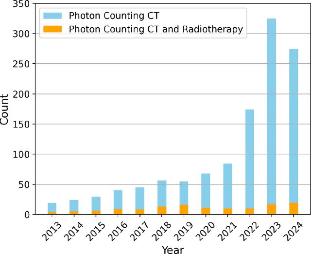
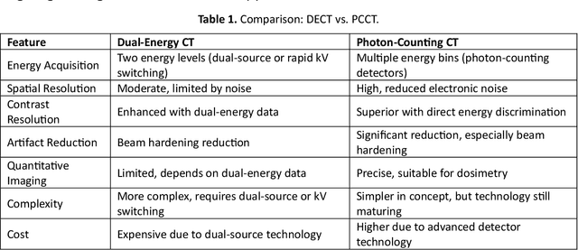


Photon-counting computed tomography (PCCT) marks a significant advancement over conventional energy-integrating detector (EID) CT systems. This review highlights PCCT's superior spatial and contrast resolution, reduced radiation dose, and multi-energy imaging capabilities, which address key challenges in radiotherapy, such as accurate tumor delineation, precise dose calculation, and treatment response monitoring. PCCT's improved anatomical clarity enhances tumor targeting while minimizing damage to surrounding healthy tissues. Additionally, metal artifact reduction (MAR) and quantitative imaging capabilities optimize workflows, enabling adaptive radiotherapy and radiomics-driven personalized treatment. Emerging clinical applications in brachytherapy and radiopharmaceutical therapy (RPT) show promising outcomes, although challenges like high costs and limited software integration remain. With advancements in artificial intelligence (AI) and dedicated radiotherapy packages, PCCT is poised to transform precision, safety, and efficacy in cancer radiotherapy, marking it as a pivotal technology for future clinical practice.