Aayush Kafle
Image Enhancement and Object Recognition for Night Vision Surveillance
Jun 10, 2020


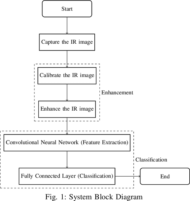
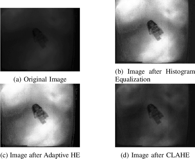
Abstract:Object recognition is a critical part of any surveillance system. It is the matter of utmost concern to identify intruders and foreign objects in the area where surveillance is done. The performance of surveillance system using the traditional camera in daylight is vastly superior as compared to night. The main problem for surveillance during the night is the objects captured by traditional cameras have low contrast against the background because of the absence of ambient light in the visible spectrum. Due to that reason, the image is taken in low light condition using an Infrared Camera and the image is enhanced to obtain an image with higher contrast using different enhancing algorithms based on the spatial domain. The enhanced image is then sent to the classification process. The classification is done by using convolutional neural network followed by a fully connected layer of neurons. The accuracy of classification after implementing different enhancement algorithms is compared in this paper.
Uncertainty Estimation in Deep 2D Echocardiography Segmentation
May 19, 2020


Abstract:2D echocardiography is the most common imaging modality for cardiovascular diseases. The portability and relatively low-cost nature of Ultrasound (US) enable the US devices needed for performing echocardiography to be made widely available. However, acquiring and interpreting cardiac US images is operator dependent, limiting its use to only places where experts are present. Recently, Deep Learning (DL) has been used in 2D echocardiography for automated view classification, and structure and function assessment. Although these recent works show promise in developing computer-guided acquisition and automated interpretation of echocardiograms, most of these methods do not model and estimate uncertainty which can be important when testing on data coming from a distribution further away from that of the training data. Uncertainty estimates can be beneficial both during the image acquisition phase (by providing real-time feedback to the operator on acquired image's quality), and during automated measurement and interpretation. The performance of uncertainty models and quantification metric may depend on the prediction task and the models being compared. Hence, to gain insight of uncertainty modelling for left ventricular segmentation from US images, we compare three ensembling based uncertainty models quantified using four different metrics (one newly proposed) on state-of-the-art baseline networks using two publicly available echocardiogram datasets. We further demonstrate how uncertainty estimation can be used to automatically reject poor quality images and improve state-of-the-art segmentation results.
 Add to Chrome
 Add to Firefox
 Add to Edge